
Author: david
The Unwritten Rules of Laboratory Life


A Joe Blogs post by Joe Roberts, PhD
When you start out in research, you’re taught the essentials: experimental design, controls, statistics, and how not to accidentally destroy expensive equipment.
What you’re not taught is that every lab, regardless of country or discipline, quietly runs on a second curriculum.
The unwritten rules.
Here are a few you only really learn by doing…
1. Experiments behave like they’re being watched
The assay that performed flawlessly for weeks will suddenly forget how to function the moment you start collecting “real” data.
Antibodies lose confidence. Cell lines become opinionated. Instruments develop personality-driven error messages.
Troubleshooting, it turns out, isn’t an occasional task, it’s a full-time relationship status.
2. You will not remember what’s in the tube
At the time, you’re certain you’ll remember exactly what “Sample 1” refers to.
Future you disagrees.
Future you is now staring at a rack of identically mysterious tubes labelled “Sample 1”, “Sample 2”, and the optimistic but unhelpful “Test”.
3. Equipment has excellent timing (and a sense of humour)
Most instruments will run perfectly for months… until a grant deadline, conference abstract, or thesis submission quietly appears on your calendar.
At that point, they become selective about functionality.
Coincidence? Possibly. Universally believed otherwise? Absolutely.
4. The Best Scientific Discussions Rarely Happen in Scheduled Meetings
Scheduled meetings are fine for alignment.
The real scientific progress tends to happen in corridors, over coffee, or while waiting for a centrifuge that still insists it has 4 minutes remaining.
Somewhere between “just thinking aloud” and “quick question”, entire projects are reshaped.
5. The manual is optional. The lab veteran is not.
Every lab has at least one person who has quietly accumulated knowledge that never made it into any official documentation.
They know which button not to press, which protocol is “technically fine but don’t tell anyone”, and how to fix things that aren’t supposed to be fixable.
Finding them early saves weeks of your life.
6. Cell culture does not respect urgency
Cells grow beautifully when you’re optimising.
The moment you scale up? They reconsider their life choices.
Experienced researchers eventually accept that biology does not respond well to deadlines or emotional bargaining.
7. Negative results are still results (eventually)
At first, negative results feel like failure.
Later, you realise they’re just data that didn’t agree with your expectations.
Some of the most useful scientific directions start with the quiet discovery that something simply doesn’t work the way you thought it would.
8. One good result is interesting. Many good results are science.
A single beautiful dataset is encouraging.
A reproducible one is convincing.
The gap between the two is where most of scientific reality lives.
9. Every Lab Has Its Own Culture
Despite performing similar research, no two laboratories operate exactly the same way.
Each develops its own traditions, routines, terminology, and preferred ways of working.
Learning a new laboratory often means learning a new culture.
10. Curiosity outlasts everything else
Techniques change. Platforms evolve. Instruments get faster and more automated.
But the driving force behind all of it stays the same: curiosity.
The best researchers don’t just follow protocols, they keep asking better questions, challenging assumptions, and exploring the unknown.
Joe’s takeaway
After working in labs across three different countries and two different hemispheres, one thing has become clear: while the science, equipment, and research questions may change, the day-to-day realities of laboratory life are surprisingly consistent.
The unwritten rules aren’t quirks to be frustrated by, they’re part of what makes experimental science what it is. They remind us that research is rarely linear, often unpredictable, and always a team effort. And more often than not, the best lessons come from experience rather than instruction manuals.
If anything, these shared experiences are a quiet reminder that wherever you are in the world, you’re part of a much larger scientific community all figuring it out together!
Until next time… happy experimenting!

On this page
- A Joe Blogs post by Joe Roberts, PhD
- 1. Experiments behave like they’re being watched
- 2. You will not remember what’s in the tube
- 3. Equipment has excellent timing (and a sense of humour)
- 4. The Best Scientific Discussions Rarely Happen in Scheduled Meetings
- 5. The manual is optional. The lab veteran is not.
- 6. Cell culture does not respect urgency
- 7. Negative results are still results (eventually)
- 8. One good result is interesting. Many good results are science.
- 9. Every Lab Has Its Own Culture
- 10. Curiosity outlasts everything else
- Joe’s takeaway
Exosome Extraction Explained
Exosome Isolation Without the Wait: Norgen Biotek's Silicon Carbide Approach
Waiting for the ultracentrifuge. Overnight incubations. Precipitation steps that compromise sample integrity. If you’re working with exosomes, you know these frustrations well, and you’ll also know how much they slow down what is otherwise one of the most productive areas of molecular biology research. Norgen Biotek’s proprietary silicon carbide (SiC) resin technology takes a fundamentally different approach to exosome isolation, one that addresses these pain points without sacrificing purity or yield.
Why Conventional Exosome Isolation Falls Short
Ultracentrifugation remains the most widely used method for exosome isolation, but its limitations are well documented. It is time-consuming, equipment-intensive, and poorly suited to high-throughput workflows. Repeated high-speed centrifugation steps can compromise vesicle integrity and co-pellet protein aggregates and lipoproteins, complicating downstream analysis. Polymer precipitation methods simplify the procedure but introduce their own purity trade-offs, and the resulting material is often incompatible with sensitive RNA or proteomic applications. For researchers working with clinical sample types such as plasma, serum and urine, where input volumes are limited and sample quality is paramount, neither approach is ideal.
The Silicon Carbide Difference: Selective Binding at the Isoelectric Point
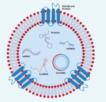
Norgen’s SiC resin technology works on a fundamentally different principle to precipitation or centrifugation-based methods. The resin selectively binds exosomes through interactions with exosomal membrane proteins at their isoelectric points under defined pH conditions. By adjusting the pH of the sample to a specific binding pH, exosomes are captured onto the SiC resin matrix via spin column chromatography. A subsequent change in pH conditions releases the intact, purified exosomes for downstream use. This means no chemical precipitation reagents, no phenol/chloroform, no protease treatments, and critically, no ultracentrifuge. The result is purified intact exosomes in the 40–200 nm size range, free from contaminating RNA-binding proteins, in under 30 minutes.

A Streamlined Workflow Across Multiple Sample Types
Norgen’s exosome purification kits support plasma, serum, urine, saliva, bacteria, and cell culture media, with kit formats optimised for each sample type. The simplified spin column workflow requires no special instrumentation or training, making it accessible regardless of your available equipment. Purified exosomes are compatible with NanoSight nanoparticle tracking analysis, transmission electron microscopy, functional assays, and direct downstream RNA isolation, without any additional clean-up steps.
For cell culture media workflows, it is worth noting that standard FBS contains substantial quantities of bovine-derived exosomes. Norgen also supplies FBS Exosome Depletion Kits, allowing researchers to prepare exosome-depleted FBS prior to use as a growth supplement – an important step for ensuring that downstream exosomal cargo analysis reflects the biology of your cells of interest rather than contaminating bovine vesicles.
For researchers looking for ready-to-use exosome material, Norgen also offers purified exosomes from human adipose-derived MSCs, suitable for use as a positive control, reference material, or workflow comparison sample.
From Exosomes to Exosomal RNA in a Single Workflow
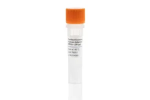
One of the most practically valuable aspects of the Norgen platform is the availability of combined purification and RNA isolation kits, which allow researchers to move from raw sample to isolated exosomal RNA in a single, uninterrupted workflow. The RNA isolation step uses Norgen’s SiC resin to capture RNA of all sizes – including microRNA – irrespective of size or GC content, without bias. The isolated RNA is free from protein-bound circulating RNA and is suitable for RT-qPCR, small RNA sequencing, transcriptomics, and biomarker discovery workflows. For plasma and serum samples where maximum purity is required, the EXTRAClean range provides an additional level of clean-up optimised for liquid biopsy and sensitive downstream assays.
The Norgen Exosome Portfolio
The full range covers intact exosome purification, combined exosome purification and RNA isolation, standalone exosomal RNA isolation from previously purified exosomes, free-circulating RNA purification, FBS exosome depletion, and bacterial extracellular vesicle isolation – with kit formats available across a range of sample volumes and throughput requirements.

Key Takeaways
- Norgen’s SiC resin selectively captures exosomes at exosomal membrane protein isoelectric points under defined pH conditions – no ultracentrifugation, precipitation reagents, or special equipment required
- Intact exosomes in the 40–200 nm size range are purified from plasma, serum, urine, saliva, or cell culture media in under 30 minutes
- Combined purification and RNA isolation kits support end-to-end workflows from sample to sequencing-ready RNA
- All sizes of exosomal RNA, including microRNA, are captured without size or GC bias
- FBS exosome depletion kits and bacterial EV isolation round out a portfolio covering the full range of exosome research needs
Explore Norgen Exosome Solutions Through Millennium Science
Millennium Science is the authorised distributor of Norgen Biotek products for researchers in Australia and New Zealand. The full Norgen exosome portfolio is available to order now.
Visit the Norgen Biotek page on the Millennium Science website or contact the team to discuss the right workflow for your sample type and application.
On this page
- Exosome Isolation Without the Wait: Norgen Biotek's Silicon Carbide Approach
- Why Conventional Exosome Isolation Falls Short
- The Silicon Carbide Difference: Selective Binding at the Isoelectric Point
- A Streamlined Workflow Across Multiple Sample Types
- From Exosomes to Exosomal RNA in a Single Workflow
- The Norgen Exosome Portfolio
- Key Takeaways
- Explore Norgen Exosome Solutions Through Millennium Science
How the Dharmacon Edit-R Platform Improves CRISPR Knockout Workflows
What’s New in the Dharmacon™ Edit-R™ CRISPR Platform

Guide RNA quality and Cas9 delivery format are two of the most important variables in any CRISPR knockout experiment. Poor guide design can reduce knockout efficiency or introduce unwanted off-target effects, while the wrong Cas9 delivery strategy can add unnecessary complexity to an already sensitive workflow.
The Dharmacon™ Edit-R™ CRISPR-Cas9 platform, available in Australia and New Zealand through Millennium Science, is designed to support reliable functional knockout experiments across a range of cell models and delivery approaches.
Two significant updates make the platform especially relevant for researchers planning CRISPR knockout workflows in 2026: predesigned human guide RNAs have been realigned to the latest RefSeq database, and a new Cas9 Protein Hybrid NLS format is now available for DNA-free RNP editing.
Designed for functional protein knockout
Not all CRISPR guide RNAs are designed with the same experimental endpoint in mind. While some guide design approaches focus primarily on generating indels at the DNA level, many knockout experiments depend on achieving functional disruption at the protein level.

The Dharmacon Edit-R algorithm has been developed and empirically validated to prioritise guides that generate functional protein knockout. Each predesigned guide RNA is scored for two key factors:
- Functionality: the likelihood of producing protein-level knockout
- Specificity: the predicted risk of off-target editing
This gives researchers a practical way to balance knockout performance and specificity depending on their cell model, target gene and downstream assay.
Guide RNA formats to match different CRISPR workflows
The Edit-R platform supports several guide RNA formats, allowing researchers to choose a workflow that suits their experimental setup rather than forcing every project into the same delivery strategy.
Synthetic crRNA system
The synthetic crRNA workflow uses three core components:
- A source of Cas9 nuclease
- A gene-specific Edit-R crRNA
- The Edit-R tracrRNA scaffold
The tracrRNA complexes with the gene-specific crRNA to direct Cas9 to the target site. This cloning-free approach helps researchers move from design to editing quickly, without needing vector construction or sequencing confirmation before starting an experiment.
This format is well suited to researchers who want a flexible, modular workflow and the ability to pair synthetic guide RNAs with different Cas9 delivery options, including purified protein, mRNA or lentiviral Cas9 expression.
Synthetic sgRNA
For researchers who prefer a single-guide format, Edit-R synthetic sgRNA combines the crRNA and tracrRNA sequences into one molecule. This simplifies handling and reduces the number of components in the workflow while retaining compatibility with DNA-free Cas9 delivery strategies.
Synthetic sgRNA can be a useful option when workflow simplicity, transfection consistency and rapid experimental setup are priorities.
Lentiviral sgRNA vectors
For stable, long-term expression, particularly in difficult-to-transfect cells or for follow-up validation after pooled screening, Edit-R lentiviral sgRNA vectors provide a more durable delivery approach.
The Edit-R lentiviral sgRNA vector expresses the gene-specific crRNA and tracrRNA as a chimeric single guide RNA under the control of a human U6 promoter. A puromycin resistance marker, driven from the mouse CMV promoter, is co-expressed in the same vector. This allows researchers to select cells carrying the integrated sgRNA construct.
This format is especially useful for:
- Difficult-to-transfect cell types
- Stable knockout studies
- Follow-up validation from pooled CRISPR screens
- Experiments requiring longer-term guide RNA expression
- Workflows where antibiotic selection is useful for enriching edited populations
All-in-one lentiviral sgRNA
The all-in-one lentiviral sgRNA format takes the stable expression workflow one step further by combining Cas9 and sgRNA expression in a single vector. This reduces the number of transduction steps required and can simplify workflow optimisation, particularly where introducing separate Cas9 and guide RNA components would add time or variability.
For researchers working in challenging cell models, the all-in-one format may provide a more streamlined route to stable CRISPR knockout experiments.
Human guide RNAs realigned to the latest RefSeq database
In late 2025, all predesigned human Edit-R synthetic and lentiviral guide RNAs were realigned against the latest NCBI RefSeq database.
This update recalculates functionality and specificity scores using the current genomic reference. Any designs that no longer met the required scoring threshold were replaced with higher-scoring alternatives.
For most genes, previously used guides remain unchanged. Where a guide has been retired, the replacement reflects a stronger predicted balance of functionality and specificity based on updated genome annotations.
Researchers who have previously ordered Edit-R guide RNAs can check specific catalogue numbers through the Horizon Discovery website. This is useful if you are repeating an earlier experiment, comparing results across projects or planning follow-up work based on a previously ordered guide.
New Cas9 Protein Hybrid NLS for DNA-free RNP editing
The new Edit-R Cas9 Protein Hybrid NLS is a purified Cas9 nuclease protein featuring an enhanced hybrid nuclear localisation signal composition. It is designed to improve nuclear delivery compared with traditional single-NLS formats.
The protein is optimised for co-electroporation or co-transfection with Edit-R synthetic guide RNAs to form ribonucleoprotein complexes, commonly referred to as RNPs. This enables a completely DNA-free CRISPR editing workflow.
DNA-free RNP editing can offer several practical advantages:
- No plasmid DNA is introduced into the system
- The risk of plasmid integration is avoided
- Promoter compatibility issues are removed
- Cas9 exposure is transient, helping to reduce off-target exposure
- The workflow can be useful for sensitive or clinically relevant cell models
Functional knockout has been demonstrated in primary human CD4+ T cells by nucleofection, and in U2OS cells using both nucleofection and lipid transfection.
The Edit-R Cas9 Protein Hybrid NLS is available in sizes from 50 µg to 5 × 500 µg, with bulk quantities available on request.
Where CRISPR knockout workflows can be applied
For molecular biology researchers, the value of these updates is not that CRISPR is new. It is that more current guide design and more flexible Cas9 delivery options can help improve the reliability of familiar workflows.
Edit-R reagents can support applications such as:
- Functional genomics studies
- Disease modelling
- Pathway analysis
- Target identification and validation
- Pooled screen follow-up
- Primary cell editing
- Cell and gene therapy research workflows
By choosing the appropriate guide RNA format and Cas9 delivery strategy, researchers can better tailor CRISPR knockout workflows to their cell type, experimental timeline and downstream readout.
Edit-R performance guarantee
Every predesigned Edit-R synthetic crRNA, sgRNA, lentiviral sgRNA and all-in-one lentiviral sgRNA is backed by an editing guarantee.
If a predesigned guide does not edit the target site when used as recommended, a replacement guide of the same format and quantity will be provided at no cost.
Key takeaways
The latest Dharmacon Edit-R updates provide researchers with a more current and flexible platform for CRISPR knockout experiments.
- Edit-R guide RNAs are designed for functional protein knockout, not just indel generation
- Predesigned human guide RNAs were realigned to the latest RefSeq database in late 2025
- Multiple guide RNA formats support synthetic, lentiviral and all-in-one CRISPR workflows
- Lentiviral formats provide stable expression options for difficult-to-transfect cells and longer-term studies
- The new Cas9 Protein Hybrid NLS supports DNA-free RNP editing with enhanced nuclear delivery
- Edit-R predesigned guide RNAs are backed by an editing guarantee
Explore Dharmacon Edit-R CRISPR solutions
Millennium Science supplies Revvity’s Dharmacon Edit-R CRISPR platform across Australia and New Zealand, supporting researchers with genome editing reagents, workflow selection and technical guidance.
Interested in learning more?
- Request pricing for Edit-R CRISPR reagents
- Discuss the best workflow for your cell model
- Explore DNA-free RNP editing strategies
- Speak with a CRISPR product specialist
On this page
- What’s New in the Dharmacon™ Edit-R™ CRISPR Platform
- Designed for functional protein knockout
- Guide RNA formats to match different CRISPR workflows
- Human guide RNAs realigned to the latest RefSeq database
- New Cas9 Protein Hybrid NLS for DNA-free RNP editing
- Where CRISPR knockout workflows can be applied
- Edit-R performance guarantee
- Key takeaways
- Explore Dharmacon Edit-R CRISPR solutions
From Samples to Insight: Meet the New Agilent Cytation 9
A Joe Blogs post by Joe Roberts, PhD
As biological models become more complex – from 3D cultures to longitudinal live-cell assays – the real bottleneck in research isn’t data generation. It’s integration. Imaging, quantitation, kinetics… too often these sit in separate workflows, separate instruments, and separate datasets.
The newly released Agilent Cytation 9 Cell Imaging Microplate Reader from Agilent Technologies takes a different approach: one platform, multiple data dimensions, and a clear path from samples to actionable insight.
Agilent Cytation 9: One Platform, Two Microscopes, Endless Applications

At the heart of Cytation 9 is something genuinely distinctive — it combines multimode plate reading with both inverted and upright microscopy in a single system.
This dual-microscope configuration opens up a broader experimental space:
- Inverted imaging for standard cell-based assays
- Upright imaging for applications like slides, ELISpot, or tissue sections
- Seamless switching between modalities without moving samples
It’s not just consolidation – it’s capability expansion.
Designed to Extract More Data Per Experiment
Cytation 9 is clearly built around maximising information density.
Key enhancements include:
- Expanded image cube capacity to capture more fluorescence channels per well
- High-resolution APO objectives for improved image quality
- Wide range of imaging modes, including fluorescence, brightfield, phase contrast, and multi-colour imaging
The result? Richer datasets from every experiment – without increasing sample volume or complexity.
Speed and Throughput Without Compromise
One of the most practical upgrades is throughput.
Cytation 9 delivers:
- Faster imaging workflows to move from acquisition to analysis more efficiently
- Automated features like laser autofocus and montage imaging
- Support for high-density formats (up to 1536-well plates)
Combined, these improvements help labs scale from routine assays to higher-throughput screening without rethinking their entire workflow.

Built for Live-Cell and Kinetic Workflows
Where Cytation 9 really comes into its own is in dynamic biology.
With software-controlled environmental regulation, including CO₂ and O₂ control, temperature, and humidity, the system enables stable long-term live-cell imaging.
This allows researchers to:
- Track cellular responses over time
- Run kinetic assays with confidence
- Reduce variability introduced by external incubation systems
It’s a shift from static snapshots to continuous biological insight.
Flexibility That Grows With Your Lab
Cytation 9 isn’t a fixed system – it’s modular.
You can:
- Add imaging modes as your applications evolve
- Integrate with automation platforms like BioSpa™ or BioStack™
- Work across diverse labware, from plates to flasks and slides
This makes it equally relevant for core facilities, translational labs, and drug discovery environments where needs change rapidly.
Joe’s Takeaway
The Cytation 9 stands out because it doesn’t force a trade-off between imaging and quantitation – it integrates both in a way that actually improves experimental design.
The addition of upright microscopy alongside the traditional inverted setup is a particularly smart move. It broadens what the system can do without adding complexity, and that’s ultimately what most labs need: more capability, not more workflow friction.
If your work involves cell-based assays, imaging, or high-content workflows, Cytation 9 is well worth a closer look.
If you’re interested in a demo or exploring how this could fit into your lab, feel free to contact us today.
Until next time… happy experimenting!
On this page
Why IFN-γ Remains One of the Most Measured Cytokines in Immunology Research
A Joe Blogs post by Joe Roberts, PhD
If you spend any time studying immune responses, you’ll quickly encounter interferon gamma (IFN-γ). It’s one of the most widely measured cytokines in immunology research, and at Millennium Science, it’s also one of the most frequently ordered reagents in our catalogue.
That’s hardly surprising. IFN-γ sits at the centre of many immune processes and is widely used as a functional readout of cellular immune activity. From infectious disease research to cancer immunology and vaccine development, measuring IFN-γ often provides a direct window into how immune cells are responding.
Let’s take a closer look at why this cytokine is so important, and the different ways researchers are measuring it today.
What is IFN-γ?
Interferon-γ (IFN-γ) is a type II interferon primarily produced by:
- CD4⁺ Th1 cells
- CD8⁺ cytotoxic T cells
- Natural killer (NK) cells
Unlike type I interferons (such as IFN-α and IFN-β), which are produced by many cell types, IFN-γ is mainly produced by activated immune cells during adaptive and innate immune responses.
Functionally, IFN-γ contributes to several important immune processes:
- Antiviral immunity – enhances immune responses against infected cells
- Immunomodulation – upregulates antigen presentation through increased MHC expression
- Macrophage activation – a key signal driving classical macrophage activation
- Pro-inflammatory signalling – promotes Th1-type immune responses
Because of these roles, IFN-γ is frequently used as a readout of cellular immune activation, particularly when studying T-cell responses.
Why Do Researchers Measure IFN-γ So Often?
In many immune assays, IFN-γ acts as a functional marker of antigen-specific immune activity.
Researchers often measure IFN-γ to:
- Evaluate vaccine-induced immune responses
- Assess T-cell immunity to infection
- Study tumour-specific immune responses
- Characterise Th1-biased immune signalling
- Monitor cell therapy potency
This helps explain why IFN-γ assays remain among the most requested cytokine reagents in our portfolio.
Mabtech Offers Four Ways to Measure IFN-γ
Different experiments require different levels of sensitivity, multiplexing, or single-cell resolution. Fortunately, IFN-γ can be detected using several complementary technologies.
- ELISA – Quantifying Secreted IFN-γ
ELISA remains one of the most widely used cytokine detection methods.
It provides:
- Quantitative measurement of IFN-γ in supernatants, serum, or plasma
- High sensitivity and reproducibility
- A straightforward workflow compatible with most labs
ELISA is ideal when you want to measure total cytokine secretion from a population of cells.
- ELISpot – Detecting IFN-γ at the Single-Cell Level
ELISpot takes cytokine detection one step further by measuring secretion from individual cells.
This technique enables researchers to:
- Quantify IFN-γ-secreting cells
- Detect rare antigen-specific T cells
- Achieve exceptional sensitivity
Because of this sensitivity, ELISpot has become a widely used approach for T-cell immune monitoring, particularly in vaccine and immunotherapy research.
- FluoroSpot – Multiplex Cytokine Detection
FluoroSpot expands on ELISpot by allowing simultaneous detection of multiple cytokines using fluorescent detection.
This makes it possible to identify polyfunctional immune cells, which can provide deeper insights into immune responses.
For example, researchers can detect:
- Triple-secreting IFN-γ / IL-2 / TNF-α T cells
- Killer cells secreting IFN-γ and Granzyme B
This type of analysis provides a much richer picture of immune responses, especially when studying complex cell populations.
- EYRAPlex – Multiplex Cytokine Quantification
For researchers who want high-throughput multiplex cytokine profiling, EYRAPlex assays provide a powerful alternative.
EYRAPlex enables:
- Simultaneous measurement of multiple cytokines
- High-throughput cytokine profiling
- Compatibility with the EYRA instrument platform and most Flow Cytometers
This approach is ideal when researchers want to explore complex cytokine signatures rather than focusing on a single immune marker.
EOFY Promotion: 10% Off IFN-γ FluoroSpot Assays
Because IFN-γ remains such a critical immune readout, we’re currently running a limited EOFY promotion.
Until 30 June, you can receive: 10% off Mabtech IFN-γ FluoroSpot assays
If you’re already measuring IFN-γ responses, or planning a new T-cell study, it’s a great opportunity to expand your toolkit.
Final Thoughts
From infection biology to immunotherapy, IFN-γ remains one of the most informative cytokines to measure when studying cellular immunity.
Whether you’re quantifying cytokine secretion with ELISA, detecting rare antigen-specific cells with ELISpot, studying polyfunctional responses with FluoroSpot, or analysing complex cytokine networks with EYRAPlex, there are now multiple powerful tools available to explore IFN-γ biology.
If you’d like more information about IFN-γ assays or the current EOFY promotion, feel free to reach out.
Until next time… happy experimenting!
Seeing More, Doing More: Agilent’s BioTek Imaging Portfolio in Action!
A Joe Blogs post by Joe Roberts, PhD
Modern life science research increasingly demands more information from fewer samples, delivered faster and with greater confidence. Whether you’re studying immune cell function, complex 3D models, or subtle phenotypic changes, imaging is central to generating meaningful insights. Agilent’s BioTek imaging portfolio is designed to meet this challenge, combining high-quality microscopy, flexible multimode detection, and live-cell capability into integrated platforms that scale with your research needs.
Cytation and Lionheart: Imaging Platforms That Scale With Your Research
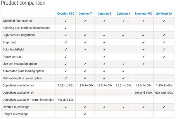
The Agilent BioTek Cytation™ range spans entry-level widefield imaging through to advanced confocal workflows, combining automated microscopy with multimode microplate reading in a single, integrated platform. Designed to scale with experimental complexity, Cytation systems support routine plate-based assays, high-content imaging, and live-cell experiments with environmental control, making them suitable for laboratories of all sizes and budgets.
For labs requiring dedicated automated microscopy, the Agilent BioTek Lionheart™ FX and LX microscopes deliver high-performance imaging in a compact footprint. These systems support a wide range of objectives and imaging modalities, with robust autofocus, flexible incubation options, and automation-ready workflows. Lionheart platforms are particularly well suited to phenotypic profiling, kinetic assays, and live-cell imaging where precision and repeatability are essential.
A detailed product comparison table below highlights the specific capabilities of each Cytation and Lionheart model.
Why Agilent BioTek Imaging Stands Out
Across the portfolio, Agilent imaging platforms share several key advantages:
- Support for 6- to 1536-well plates, slides, dishes, and flasks.
- Compatibility with automation solutions like BioStack™, BioSpa™, and BenchCel™.
- Live-cell imaging with CO₂/O₂ control, shaking, and temperature incubation.
- Advanced imaging capabilities: Z-stacking, Z-projection, montage stitching, and time-lapse videos.
- Hybrid optics for sensitive and specific fluorescence detection.
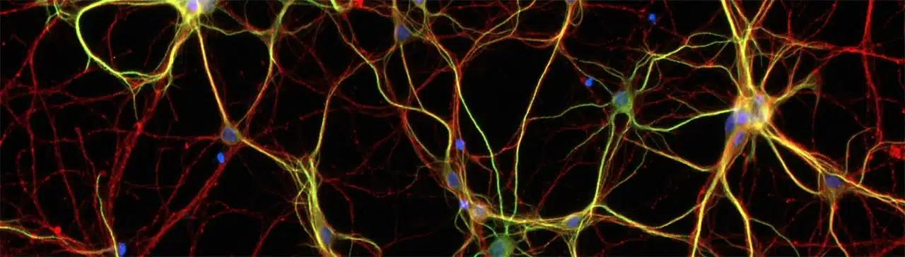
Applications include wound healing assays, cell migration and invasion studies, immunofluorescence, neurite outgrowth analysis, 3D cell models, and ADME/Tox experiments. By unifying imaging and detection in a single platform, Agilent BioTek systems help labs move beyond single-endpoint measurements to richer, biologically meaningful data.
Joe’s Takeaway
Whether you’re starting with simple plate-based assays or pushing the boundaries of confocal or automated microscopy, the Agilent BioTek imaging portfolio gives you the flexibility, performance, and scalability to see more and do more.
If you’re interested in a demo, contact us today!
Until next time… happy experimenting!
One Year In: Reflections on Life, Science and Supporting Discovery at Millennium Science
A Joe Blogs post by Joe Roberts, PhD
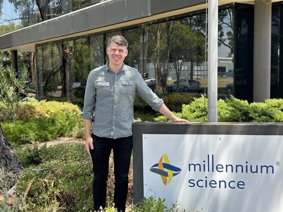
It’s hard to believe it’s already been one year since I joined Millennium Science. Over the past 12 months, I’ve had the privilege of working closely with researchers across Australia and New Zealand, supporting everything from early-stage assay development through to established, high-throughput workflows. It’s been a year full of learning, connection, and a renewed appreciation for the science happening right on our doorstep.
A Front-Row Seat to Great Science
One of the biggest positives of this role has been the variety. No two weeks look the same, from visiting labs and running demos, to attending conferences and catching up with researchers over morning teas. Those conversations, whether at the bench or over a coffee, often provide the most valuable insights into what researchers actually need.
Events like CYTO-Connect and ASI have been particular highlights. There’s something energising about seeing the community come together, sharing data, debating approaches, and learning from one another. Being able to support these meetings, demonstrate new technologies, and have real, practical conversations about workflows has been incredibly rewarding.
From the Bench to the Field
Coming into this role from a background as a Post-Doctoral Research Fellow, I initially saw the move as an opportunity to apply my scientific training in a different way. What I’ve learned is just how valuable that bench-side experience is when supporting researchers, understanding experimental constraints, timelines, and the realities of generating reliable data.
Over the past year, that experience has shaped how I work: launching and writing Joe Blogs to share practical insights from the field, hosting webinars to break down new technologies, providing hands-on instrument and assay demos, and presenting at national conferences. Across all of these, the goal has remained the same, to translate complex technologies into something genuinely useful for real research questions.
What the First Year Has Taught Me
This past year has also been a steep (and enjoyable) learning curve. A few key takeaways stand out:
- No two labs are the same – listening is always more valuable than assuming.
- Technology is only powerful when it’s accessible – training, support, and context matter just as much as specifications.
- Collaboration drives outcomes – the best results come from strong partnerships between researchers, suppliers, and local support teams.
Science Is Serious – But Culture Matters Too
At Millennium Science, science is serious – but culture matters just as much. It’s not just about cutting-edge tech; it’s about curiosity, collaboration, and actually enjoying the workday. We’ve got a dog-friendly office, share knowledge across teams, and even throw epic end-of-year battle cart competitions (which Joe Blogs won last year).
That mix of expertise, energy, and a little friendly fun makes it a joy to help researchers tackle real-world challenges, turning complex tech into solutions that actually make a difference in the lab.
Looking Ahead
As I head into year two, I’m excited to continue supporting researchers through lab visits, demos, webinars, conferences, and everything in between. If you’re planning a new project, exploring a new technology, or just want to talk through an idea over a coffee, I’d love to hear from you (contact us today).

Until next time… happy experimenting!
Common Challenges in RNA Extraction and How to Solve Them
From Sample to Data: Cleaner RNA, Better Results
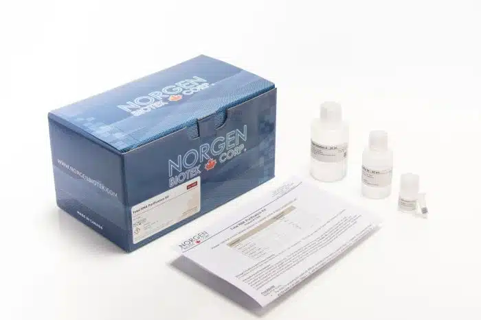
RNA extraction is a crucial step in molecular biology, but it often comes with obstacles that can impact downstream applications such as RT-qPCR, sequencing, and microRNA analysis. Researchers in Australia and New Zealand frequently face challenges with low yield, degraded RNA, or inhibitor contamination. Fortunately, Norgen Biotek’s Total RNA Purification Kits, distributed by Millennium Science, offer reliable and comprehensive solutions.
Challenge 1: Low RNA Yield
Working with small or precious samples often results in low RNA recovery.
Solution: Norgen’s Total RNA Purification Kits are optimised to maximise yield across a wide range of sample types, including blood, plasma, tissues, and cultured cells. With just one kit, researchers can extract total RNA covering all sizes from large mRNA down to small RNA species such as microRNA ensuring nothing important is lost.
Challenge 2: RNA Degradation
RNA is notoriously unstable and highly susceptible to degradation during purification.
Solution: Norgen’s patented silicon carbide (SiC) resin technology protects RNA integrity throughout the process. This unique chemistry binds and isolates all RNA species including fragile small RNAs without the need for harsh chemicals like phenol, chloroform, or alcohol precipitation. By operating under gentler conditions than traditional silica methods, the SiC resin preserves RNA integrity and delivers intact RNA ready for sensitive applications such as sequencing, RT-qPCR, and microRNA profiling.
Challenge 3: Presence of Inhibitors
Contaminants such as heme, salts, or proteins can inhibit enzymatic reactions and compromise results.
Solution: Norgen kits efficiently remove inhibitors during the purification process, providing RNA that is pure, stable, and immediately suitable for downstream workflows, from gene expression analysis to diagnostic assay development.
Take the uncertainty out of RNA extraction. Request a quote or contact Millennium Science today to learn more about Norgen Biotek’s complete range of RNA purification solutions.
Why Choose Norgen Biotek with Millennium Science
As the exclusive distributor of Norgen Biotek products in Australia and New Zealand, Millennium Science, based in Melbourne, provides researchers with responsive technical support, and competitive pricing. By combining innovative technology with local expertise, Millennium Science ensures scientists have the tools they need to achieve reproducible, high-quality results.
Practical Tips for Reliable cDNA Synthesis in qPCR Workflows
Why cDNA synthesis quality matters for qPCR results
In any qPCR workflow, the quality of your cDNA is critical. Poor reverse transcription can lead to low sensitivity, biased results, and unreliable gene expression data. By following a few best practices and using a high-performance kit such as the SensiFAST™ cDNA Synthesis Kit from Meridian Bioscience (Bioline), together with Meridian’s RNA Isolation Kits, researchers in Australia and New Zealand can achieve consistent, reproducible results from sample to data.

Tip 1: Start with High-Quality RNA
The integrity of your RNA will directly impact your cDNA synthesis. Meridian’s RNA Isolation Kits provide high-yield, inhibitor-free RNA suitable for sensitive downstream applications. Whether you are working with blood, tissues, or cultured cells, these kits deliver clean RNA that sets the foundation for reliable cDNA synthesis.
Tip 2: Choose a Kit Designed for Full Coverage
Not all cDNA synthesis kits are created equal. Some introduce a 3′ bias, leaving long transcripts under-represented. The SensiFAST cDNA Synthesis Kit uses a combination of anchored oligo(dT) primers (which ensure the 3′ ends of mRNAs are always captured) and random hexamer primers (which prime across the whole transcript). This ensures a more accurate reflection of gene expression.
Tip 3: Optimise for Low Input Samples
When working with small or precious samples such as biopsies, sensitivity matters. The SensiFAST kit can generate high-quality cDNA from as little as 1 picogram of RNA, making it ideal for low-input applications. Pairing it with Meridian’s RNA Isolation Kits ensures even small amounts of RNA are recovered efficiently and protected from inhibitors.
Tip 4: Validate with a Trial Kit
For labs trialling Bioline solutions for the first time, trial kits are available. These smaller packs let you test both the RNA Isolation Kit and the SensiFAST cDNA Synthesis Kit in your own workflows before scaling up—helping you validate results with minimal upfront cost.
Why Choose Millennium Science
As the exclusive distributor of Meridian Bioscience Bioline products in Australia and New Zealand, Millennium Science provides:
- Local stock availability for faster delivery
- Expert technical support across RNA isolation and cDNA synthesis workflows
- Competitive pricing and access to trial kits for first-time users
From RNA isolation to first-strand synthesis, every step matters in your qPCR workflow. By combining Meridian’s RNA Isolation Kits with the SensiFAST cDNA Synthesis Kit, researchers can achieve reproducible, high-quality results, even with challenging or low-input samples.
The Reagent Insider: Advancing Alzheimer’s research
Tips, insights and updates
As the leading cause of dementia, Alzheimer’s disease impacts over 50 million people worldwide. In Australia, dementia has recently overtaken heart disease as the leading cause of death, particularly among women. While current treatments focus on symptoms, new research aims to target the underlying disease from clearing amyloid-β, inhibiting tau aggregation, and modulating neuroinflammation to understanding the gut-brain axis and early metabolic changes in AD.
Hot Product
StressMarq’s high-quality tau, amyloid-β, and alpha-synuclein proteins support research into the mechanisms underlying Alzheimer’s disease. Contact us for details.

Exploring the Gut-Brain Link in Alzheimer’s
Emerging studies show gut health influences cognitive function, with a balanced microbiome and Mediterranean diet potentially lowering dementia risk. Using Norgen Biotek’s Stool Nucleic Acid Collection and Preservation Systems, researchers are uncovering novel approaches to tackle cognitive decline.
Connect with Narisa Dawar to learn more about Norgen Biotek.
Lab Favourite
Silence Alzheimer’s-related genes with siRNA.
Targeting tau reveals its role in microtubule stability and neurofibrillary tangles. Check out Revvity’s Dharmacon™ siRNA solutions.


MedChemExpress Neurodegenerative Disease-related Compound Library
This library of 2,960 compounds targets amyloid-β, dopamine receptors, COMT, LRRK2, 5-HT receptors, and more! Perfect for high-throughput, high-content screening of neurodegenerative mechanisms and potential treatments for Alzheimer’s, Parkinson’s, and MND.
Curious about MCE’s compound libraries? Christine Goy is just an email away!

Protocol Tip
Stay on track in Alzheimer’s research. Use InvivoGen’s Mycoplasma Strip to detect contamination early to ensure reliable neuronal and glial culture results. Email Christine Goy for details.


Metabolomics and the Brain
Understanding early metabolic changes in Alzheimer’s disease may identify new therapeutic targets and predictive biomarkers. Metabolomics can detect these subtle biological shifts before clinical symptoms appear, supporting the development of early interventions and treatments that may improve outcomes for people with AD.

Mabtech IRIS 2 in Immunology Research: Insights from Dr Liam Kealy
Introduction
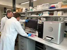
When accuracy, sensitivity, and throughput matter, immunologists are turning to advanced platforms like the Mabtech IRIS 2. Dr. Liam Kealy, Postdoctoral Researcher at Monash University, recently shared his insights into how the IRIS 2 elevated his work on plasma cells and antibody secretion and his experience working with Millennium Science. His testimonial highlights why the IRIS 2 is considered a gold standard for ELISpot, FluoroSpot, and viral focus-formation assays.

The Research Challenge: Studying Plasma Cells and Antibodies
Before adopting the Mabtech IRIS 2, Dr. Kealy and his team faced a significant challenge: the need for a multiplex approach to directly study plasma cells and the diverse antibodies they secrete. Conventional workflows lacked the flexibility and precision required for such complex immunology studies.
Gold-Standard Accuracy in Spot Detection
The Mabtech IRIS 2 solved this challenge by providing high-throughput, accurate, and user-friendly spot detection. According to Dr. Kealy:
“The IRIS 2 by Mabtech stands as the gold standard in spot detection and analysis. Equipped with advanced software, it enables high-throughput spot counting with unparalleled accuracy. Moreover, it is capable of detecting up to four colours and can provide users with additional metrics like relative spot volume.”
Applications in Immunology
This capability has opened new possibilities for his research, including ELISpots, FluoroSpots, and viral focus-formation assays. With its ability to measure multiple analytes simultaneously, the IRIS 2 enables researchers to collect richer datasets while saving time and samples.

Advanced Analysis with the Mabtech IRIS 2
The intuitive interface and advanced software tools mean researchers can move seamlessly from data acquisition to analysis. For immunologists studying antibody secretion, vaccine responses, or infectious disease mechanisms, this efficiency translates to deeper insights and faster progress.
Experience working with Millennium Science
Equally important, the collaboration with Millennium Science has played a role in supporting Dr. Kealy’s research goals. He notes:
“I always immensely enjoy chatting science with Millennium Science and working with their team to design bespoke strategies to tackle my research questions.”

Takeaway
For researchers seeking accuracy, flexibility, and high-throughput performance in multiplex immunology assays, the Mabtech IRIS 2 offers a user-friendly solution trusted by leading scientists.
Unchained Labs instruments: grant writing resources and sole-source documents
How these instruments strengthen your funding proposal
When you’re writing a capital-equipment or method-development proposal, reviewers want to see clear capability gaps and why this specific tool is required. Below is a quick guide to Unchained Labs’ key instruments and why researchers across biologics, gene therapy and nanoparticle R&D include them in funding proposals. Request the vendor sole-source document for any instrument using the form at the bottom of this page.
Aunty: micro-volume, high-throughput biologics stability
Why include it in your proposal
- Measures Tm, Tagg and Tsize in a single 1-hour run by combining full-spectrum fluorescence with SLS and DLS (single instrument).
- 96-well sealed quartz plate, only 8 µL per well; ideal for large screens and long-term/isothermal studies.
- AAV/viral vector stability workflow tracks genome ejection, capsid disruption and aggregation.
- Unmatched speed: Aunty can measure size and fluorescene for a full 96-well plate of samples every minute.

Stunner: UV/Vis + (R)DLS in one platform
Why include it
- Only system to combine UV/Vis with rotating-angle DLS (RADLS) so you get concentration, size and polydispersity from the same 2 µL sample.
- For AAV, reports total capsid, full/empty ratio, size and aggregation in one workflow. For LNPs, quantifies payload by UV/Vis (Unmix) while measuring particle metrics by RADLS.
- 96 samples/run, automation-friendly microfluidic plates; wide absorbance range (0.03-275 OD10mm).

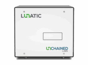
Lunatic: 96-well, 2 µL microvolume QC (DNA/RNA/protein)
Why include it
- 5 minutes for 96 samples (typical) and no drop-and-wipe – microfluidic plates handle sample loading and retention.
- UV/Vis plus Unmix applications to quantify targets and common impurities across a very wide linear range (no serial dilutions).
- Automation-friendly; GLP options available.
Leprechaun: single-particle LV & EV characterisation (SP-IRIS + fluorescence)
Why include it
- Combines SP-IRIS with fluorescence microscopy to measure concentration, size and protein composition at the single-particle level.
- LV assays quantify capsid and RNA-containing particles, empty capsids, aggregates, soluble p24, and EV contaminants from the same sample – minimal prep.
- Exosomes down to 35 nm, with phenotyping across up to 4 markers at once; 16 samples/run; 1-25 µL per sample.

Sunscreen: high-throughput nanoparticle formulation screening
Why include it
- Automates 96 unique formulations <6 hours with flexible TFR/FRR and optional inline dilution – walk-away runs cut time and material use.
- Uses reusable glass microfluidic “Sunnies” (40-200 nm applications) to improve reproducibility and reduce consumables.
- Designed to transfer methods directly to Sunshine for process development and continuous production.

Sunshine family: process development to continuous manufacture
Why include it
- Direct method transfer from Sunscreen to scale-up: Sunshine sits downstream of Sunscreen; the same pumping technology and microfluidic Sunnies are used across the Sunny Suite, so process parameters and learnings carry forward from screening into process optimisation and production.
- Built for optimisation and continuous operation: Sunshine is described as an automated process optimisation and continuous pumping system for moving to larger volumes, helping you justify end-to-end capability within one ecosystem.
- Lower implementation risk: Using the same Sunny Suite hardware and software stack across steps reduces re-validation effort when moving from hit-finding to robust production protocols.

Big Tuna: automated buffer exchange & concentration
Why include it
- Hands-off UF/DF you can trust: Big Tuna fully automates plate-based ultrafiltration/diafiltration to buffer-exchange, concentrate and clean up proteins, AAVs, LNPs and other biomolecules – cutting manual steps and setup time.
- High throughput, flexible formats: Run up to 96 samples (Unfilter 96) or 24 samples (Unfilter 24) in one experiment; the plates are designed for pressure-driven UF/DF, supporting efficient, parallel prep.
- Tight process control for reproducibility: Gentle mixing and positive pressure deliver uniform exchange; the system monitors volumes and auto-adjusts cycles to hit your target % exchange and concentration – then can reconcentrate if needed.
- Great for formulation and characterisation workflows: Ideal upstream of stability or particle analyses; Unchained Labs shows streamlined formulation screens and sample prep when pairing Big Tuna with other tools in the portfolio.

Request Sole Source document
On this page
- How these instruments strengthen your funding proposal
- Aunty: micro-volume, high-throughput biologics stability
- Stunner: UV/Vis + (R)DLS in one platform
- Lunatic: 96-well, 2 µL microvolume QC (DNA/RNA/protein)
- Leprechaun: single-particle LV & EV characterisation (SP-IRIS + fluorescence)
- Sunscreen: high-throughput nanoparticle formulation screening
- Sunshine family: process development to continuous manufacture
- Big Tuna: automated buffer exchange & concentration
- Request Sole Source document
Exploring the microbial landscape of No- and Low-Alcohol wines
Why watch
The fast-growing no- and low-alcohol (NOLO) category brings fresh microbiology challenges: dealcoholisation can affect microbial stability and sensory profile, and lower ethanol can increase spoilage risk. This concise session explores what’s changing in the microbial landscape – and what that means for QA.
What you’ll learn
- How dealcoholisation impacts microbial stability and the sensory profile of wines.
- An overview of >800 microorganisms isolated across process stages, and how DNA sequencing was used for identification.
- Insights from testing a subset for tolerance to ethanol and common antimicrobials.
- Practical strategies to manage spoilage and support product stability in NOLO winemaking.
Speaker
Andrea Bustos Jarufe is a Master’s candidate in Wine Microbiology at the University of Adelaide, working within the CRC-P “Advancement of Australian Lifestyle Wines”. Her research focuses on improving microbial stability and sensory quality in dealcoholised wines.
On this page
Looking for a distributor?
Distribute your products across Australia and New Zealand with Millennium Science
Register your interestMillennium Science partners with carefully selected manufacturers who are leaders in their chosen fields and are equally customer focused. Our product portfolio covers a wide scope of applications to offer comprehensive workflow solutions to researchers. Applications include flow cytometry, imaging, single cell genomics, spatial transcriptomics, lab automation, PCR/qPCR and many others in the molecular biology and cellular analysis.
For our business partners, we offer extensive market knowledge, expert logistics capabilities to facilitate customs clearance, a highly credentialed technical sales force with representation for all major research centres and outstanding local applications support and service.

What We Offer Our Suppliers
Our Sales Force
Our Sales Force, which covers all of Australia and New Zealand, includes a dedicated and highly experienced sales team of Account/Territory Managers, customer support, finance and logistics staff. They are backed by Product Managers and highly qualified Program Directors/Commercial Leads with specialised product line expertise, effectively targeting the appropriate customer base and offering informed technical advice to our clients.
Marketing
From traditional tradeshow participation to edgy social media engagement, we have unquestionably established the premier marketing organisation of any Australian/NZ distributor. Our broad-based activities enable us to engage the diverse research community through their preferred communication channels to educate customers about new applications and technologies on behalf of our suppliers.
Our Service Team
We pride ourselves in delivering excellent service and customer support for the premium instrumentation that our company represents. Our qualified service engineers are trained on all instruments. Along with a dedicated service contractor for Western Australia and New Zealand, our Service Team covers the full ANZ region.
A Leader in Industry Logistics
Administered by the Australian Border Force, Australian Trusted Trader reduces red tape for Trusted Traders at the border, improves certainty in export markets, and expedites the flow of their cargo in and out of Australia. This means faster access to market. We are proud to be one of the only Life Sciences distributors to have an accredited Australian Trusted Trader status to enable efficient and expedient logistical services to our customers.
A Reputation of Excellence
Millennium Science is proud that it has regularly been recognised by leading manufacturers for its outstanding commercial capabilities.
A small number of our awards include:
🏆Recognition as first ever Asia-Pacific Gold Tier distributor by 10x Genomics
🏆Two-time winner of the PacBio’s Best APAC Marketing Award
🏆Distributor of the Year, APAC – 10x Genomics
🏆Marketing of the Year, APAC – 10x Genomics
🏆Strategic Leadership and Growth Award – MedChemExpress
Register your interest
Welcome to Joe Blogs
A Fortnightly Take on Life Science Technology 🔬
Hi, I’m Joe Roberts, PhD – Product Manager here at Millennium Science, immunologist at heart, and someone who’s passionate about helping researchers get the most out of their tools, assays and experimental design. My career has taken me through PhD, post-doctoral, and senior scientist roles, including roles within flow cytometry core facilities across the UK, Ireland and Australia. That journey has shaped a deep appreciation for the technologies that enable great science.
Here at Millennium Science, our mission is to ignite life science discovery with revolutionary technology!
This blog series – which I’m calling Joe Blogs – is my way of sharing insights, practical tips, and a few behind-the-scenes stories from the technologies I work with every day. From immunoassays and cell-based analysis to real-time monitoring and flow cytometry, I’ll be unpacking the platforms and reagents that drive discovery across Australia and New Zealand’s life science community.
What You Can Expect from Joe Blogs ✍️
Whether you’re running your first experiment on a new platform or refining an advanced workflow, Joe Blogs is designed to help you:
✅ Understand what your instruments can (and can’t) do
✅ Compare assay options with clarity and context
✅ Get practical insights from someone who’s been there
✅ Stay on top of emerging techniques and technologies
Think of it as a conversation, where we talk shop about the platforms you’re using, the questions you’re asking, and the science you’re working to advance.
Joe Blogs – Upcoming Topics 📅
Here’s a taste of what’s in the pipeline:
- Spot the Difference: ELISpot vs FluoroSpot vs ELISA
- Real-Time Results: What xCELLigence Really Tells You About Cell Health
- NovoCyte or NovoCyte Opteon? Choosing the Right Flow Cytometer
- Setting Up Your First ELISpot Assay: Tips for Success from Day One
I’d Love to Hear from You 💬
Got a burning question, a technical challenge, or a topic you’d love to see unpacked in a future post?
Let me know! I’d really like this blog to reflect the real conversations and challenges happening across our local research community.
Whether it’s about assay design, data interpretation, instrument comparisons or workflow optimisation, your ideas will help shape future editions of Joe Blogs. You can reach me directly via email or through the team at Millennium Science.
Let’s Keep It Real (and Local) 🤝
As part of Millennium Science, we’re proud to partner with leading life science suppliers to bring world-class tools to Australian and New Zealand researchers, with the local support to match. My goal with Joe Blogs is to give you a bit of that support upfront: advice, clarity, and maybe even a shortcut or two.
So, whether you’re measuring T cell responses, analysing cell proliferation, or exploring new approaches to data-rich experiments, you’re in the right place.
Until next time…happy experimenting!
In the Lab with Dr Sabrina Sofia Burgener
Live Cell Imaging of Inflammasome Signalling
In this webinar, Dr Burgener shares how Agilent technologies, including the Cytation 5 Cell Imaging Multimode Reader, have advanced her inflammasome research by enabling high-throughput live-cell imaging over time – providing dynamic insights into inflammasome activation and signalling outputs.
What you’ll learn:
- The role of the NLRP3 inflammasome in immune defence and sterile inflammation
- Key mechanisms: CASP1 activation, cytokine release, gasdermin D cleavage, and pyroptosis
- Limitations of single-endpoint assays in inflammasome research
- How live-cell imaging enables temporal analysis of ASC speck formation, cytokine secretion, and cell death
- Newly established, standardised assays for tracking inflammasome activation in real-time
On this page
10x Genomics Grant Application Resources
Grant Application Resources for Single Cell and Spatial Transcriptomics
Secure funding with confidence using our comprehensive 10x Genomics grant application resources, designed to help you articulate the scientific value, feasibility, and innovation of your single‑cell and spatial biology projects. Millennium Science partners with 10x Genomics to bring you expertly curated resources that strengthen your research proposals and increase your chances of success.
Why 10x Genomics?
10x Genomics technologies are widely used in peer‑reviewed research, enabling high‑resolution single‑cell and spatial profiling across diverse biological systems. With over a decade of innovation, extensive validation, and thousands of publications demonstrating impact, these platforms help you justify the scientific significance and methodological rigour of your proposals.
Downloadable Grant Resources
Enhance your applications with ready‑to‑use materials for key 10x Genomics platforms:


What’s Included in the Resources
Our downloadable guides provide:
- Clear technology descriptions that strengthen methodological justification
- Workflow and sample preparation overviews aligned with best practice
- Performance metrics and validation data to support feasibility claims
- Data processing and analysis recommendations
- Sample and project size considerations for budgeting
- Up‑to‑date references and supporting publications
Additional Support Materials
Inform your proposal with broader context and background:
Tailored Assistance from the 10x‑perts
Need help framing your aims or justifying your technology choice for upcoming grant application? Book a project discussion with one of our specialists. Our team can help with experimental design, sample requirements, technology selection and data analysis planning, providing valuable input for competitive funding applications.