Discover how single cell spatial biology is transforming our understanding of colorectal cancer.
The Xenium platform enables researchers to profile gene expression directly within intact tissue, preserving the spatial relationships between tumour, immune and stromal cells that drive disease progression.
Why study colorectal cancer with Xenium?
Bulk sequencing averages signals across thousands of cells.
Xenium reveals individual cells within their native tissue architecture, allowing researchers to investigate:
• Tumour heterogeneity
• Immune excluded regions
• The invasive tumour front
• Cell neighbourhoods
• Spatial biomarker expression
• Tumour immune interactions
Why Xenium?
✓ Single cell, subcellular resolution• Tumour heterogeneity
✓ Spatial context preserved
✓ FFPE compatible
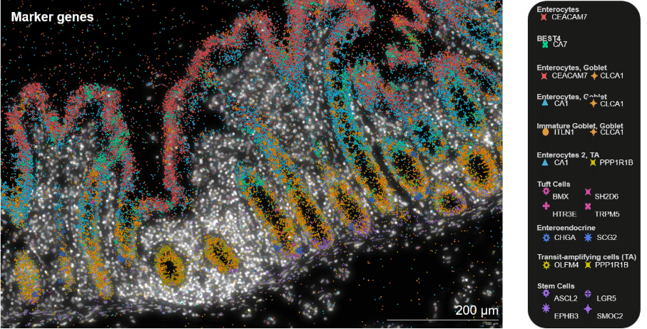
✓ Human Colon Gene Expression Panel
✓ Custom add-on panels
✓ RNA and protein profiling on the same tissue section
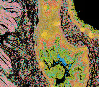
Xenium spatial transcriptomic analysis of CRC FFPE tissue. (A) RNA transcript density. (B) Multiplexed protein detection via immunofluorescence. (C) RNA clustering. (D) Post-Xenium H&E.

Formalin-fixed, paraffin-embedded (FFPE) human colorectal cancer (CRC) samples were analyzed using the Xenium Human Colon Gene Expression Panel, supplemented with 53 Immuno-Oncology–specific genes, and stained with all Xenium Protein subpanels. Over 1.7 million cells were profiled for both RNA and protein expression.