
What Are Your Cells Really Doing? Meet Agilent Cytation 1


A Joe Blogs post by Joe Roberts, PhD
What if your plate reader could also show you what your cells are actually doing?
For many cell biology experiments, a single number only tells part of the story. Fluorescence, absorbance or luminescence can give you a quantitative result, but sometimes you also want to see changes in cell morphology, number, distribution or phenotype.
The good news is that adding automated imaging to your workflow doesn’t necessarily mean investing in a large, complex or expensive imaging system.
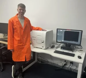
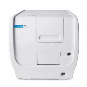
The Agilent BioTek Cytation™ 1 Cell Imaging Multimode Reader is an affordable, budget-friendly solution that combines digital microscopy with multimode plate reading, giving researchers both image-based phenotypic data and quantitative measurements from a single instrument.
And the best part? You can actually try it for yourself!
One Instrument. Imaging + Multimode Detection.
Cytation 1 is designed to make automated cell imaging accessible without requiring a dedicated, complex microscopy setup.
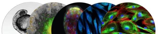
On the imaging side, Cytation 1 provides widefield fluorescence and high-contrast brightfield imaging, with magnification from 1.25x to 60x. Automated features including the XY stage, autofocus, auto exposure and LED intensity help make image capture straightforward and reproducible.
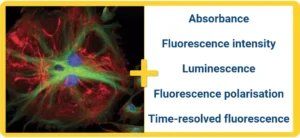
On the plate-reading side, Cytation 1 supports a range of detection modes, including fluorescence intensity, fluorescence polarisation, luminescence, time-resolved fluorescence, TR-FRET and UV-Vis absorbance.
So rather than choosing between “I need images” and “I need quantitative data”, Cytation 1 brings both into the same workflow.
What Can You Actually Do With It?
This is where Cytation 1 gets particularly interesting for cell biology.
Imagine running a cell viability or cytotoxicity assay. Your plate-reader measurement can tell you about the overall response, while imaging can provide additional information about cell number, morphology and distribution.
Or perhaps you’re studying cell proliferation or confluence. Instead of relying solely on an endpoint measurement, automated imaging can help you monitor changes across your wells over time.
Cytation 1 also supports single- and multi-colour imaging, time-lapse imaging, Z-stacking and montage imaging, opening up plenty of possibilities for cell-based research.
And if you’re working with live cells, Cytation 1 can be configured with environmental control, including temperature and CO₂/O₂ control, supporting longer-term kinetic experiments.
The platform can also accommodate a broad range of labware, from 6- to 1536-well plates, through to slides, cell culture dishes and flasks.
In other words, Cytation 1 isn’t just about taking a nice picture of your cells.
It’s about turning those images into useful quantitative data.
And What About the Software?
You don’t want to spend half your day taking images and the other half trying to work out what to do with them.
Cytation 1 uses Agilent BioTek Gen5™ software for image capture, plate reading and analysis. The workflow is designed to help automate image capture, processing and analysis, making it easier to turn images into meaningful data.
That means you can go from sample → image → analysis → data within the same platform.
Don't Just Take My Word For It — Try Cytation 1!
That’s actually the best way to find out whether Cytation 1 is right for your research.
At Millennium Science, we’re making a Cytation 1 demo unit available so researchers can get hands-on experience with the system and explore how it could work with their own applications.
Bring your questions. Bring your samples if appropriate. Let’s see what the instrument can do.
Interested in a Cytation 1 demonstration? Get in touch with us today to request a demo.
You might be surprised by just how much more information you can get from every well.
Until next time… happy experimenting!
