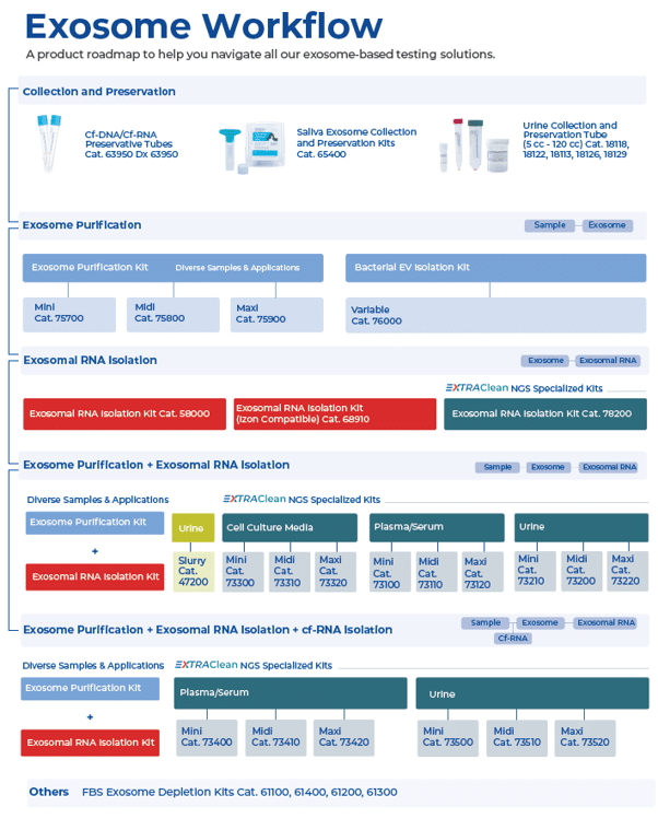
Category: Product Highlight
Exosome Extraction Explained
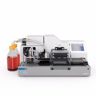
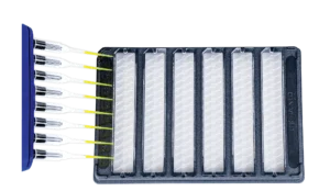
Exosome Isolation Without the Wait: Norgen Biotek's Silicon Carbide Approach
Waiting for the ultracentrifuge. Overnight incubations. Precipitation steps that compromise sample integrity. If you’re working with exosomes, you know these frustrations well, and you’ll also know how much they slow down what is otherwise one of the most productive areas of molecular biology research. Norgen Biotek’s proprietary silicon carbide (SiC) resin technology takes a fundamentally different approach to exosome isolation, one that addresses these pain points without sacrificing purity or yield.
Why Conventional Exosome Isolation Falls Short
Ultracentrifugation remains the most widely used method for exosome isolation, but its limitations are well documented. It is time-consuming, equipment-intensive, and poorly suited to high-throughput workflows. Repeated high-speed centrifugation steps can compromise vesicle integrity and co-pellet protein aggregates and lipoproteins, complicating downstream analysis. Polymer precipitation methods simplify the procedure but introduce their own purity trade-offs, and the resulting material is often incompatible with sensitive RNA or proteomic applications. For researchers working with clinical sample types such as plasma, serum and urine, where input volumes are limited and sample quality is paramount, neither approach is ideal.
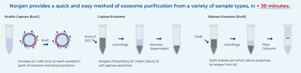
The Silicon Carbide Difference: Selective Binding at the Isoelectric Point
Norgen’s SiC resin technology works on a fundamentally different principle to precipitation or centrifugation-based methods. The resin selectively binds exosomes through interactions with exosomal membrane proteins at their isoelectric points under defined pH conditions. By adjusting the pH of the sample to a specific binding pH, exosomes are captured onto the SiC resin matrix via spin column chromatography. A subsequent change in pH conditions releases the intact, purified exosomes for downstream use. This means no chemical precipitation reagents, no phenol/chloroform, no protease treatments, and critically, no ultracentrifuge. The result is purified intact exosomes in the 40–200 nm size range, free from contaminating RNA-binding proteins, in under 30 minutes.
A Streamlined Workflow Across Multiple Sample Types
Norgen’s exosome purification kits support plasma, serum, urine, saliva, bacteria, and cell culture media, with kit formats optimised for each sample type. The simplified spin column workflow requires no special instrumentation or training, making it accessible regardless of your available equipment. Purified exosomes are compatible with NanoSight nanoparticle tracking analysis, transmission electron microscopy, functional assays, and direct downstream RNA isolation, without any additional clean-up steps.
For cell culture media workflows, it is worth noting that standard FBS contains substantial quantities of bovine-derived exosomes. Norgen also supplies FBS Exosome Depletion Kits, allowing researchers to prepare exosome-depleted FBS prior to use as a growth supplement – an important step for ensuring that downstream exosomal cargo analysis reflects the biology of your cells of interest rather than contaminating bovine vesicles.
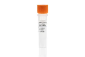
For researchers looking for ready-to-use exosome material, Norgen also offers purified exosomes from human adipose-derived MSCs, suitable for use as a positive control, reference material, or workflow comparison sample.
From Exosomes to Exosomal RNA in a Single Workflow
One of the most practically valuable aspects of the Norgen platform is the availability of combined purification and RNA isolation kits, which allow researchers to move from raw sample to isolated exosomal RNA in a single, uninterrupted workflow. The RNA isolation step uses Norgen’s SiC resin to capture RNA of all sizes – including microRNA – irrespective of size or GC content, without bias. The isolated RNA is free from protein-bound circulating RNA and is suitable for RT-qPCR, small RNA sequencing, transcriptomics, and biomarker discovery workflows. For plasma and serum samples where maximum purity is required, the EXTRAClean range provides an additional level of clean-up optimised for liquid biopsy and sensitive downstream assays.
The Norgen Exosome Portfolio
The full range covers intact exosome purification, combined exosome purification and RNA isolation, standalone exosomal RNA isolation from previously purified exosomes, free-circulating RNA purification, FBS exosome depletion, and bacterial extracellular vesicle isolation – with kit formats available across a range of sample volumes and throughput requirements.
Key Takeaways
- Norgen’s SiC resin selectively captures exosomes at exosomal membrane protein isoelectric points under defined pH conditions – no ultracentrifugation, precipitation reagents, or special equipment required
- Intact exosomes in the 40–200 nm size range are purified from plasma, serum, urine, saliva, or cell culture media in under 30 minutes
- Combined purification and RNA isolation kits support end-to-end workflows from sample to sequencing-ready RNA
- All sizes of exosomal RNA, including microRNA, are captured without size or GC bias
- FBS exosome depletion kits and bacterial EV isolation round out a portfolio covering the full range of exosome research needs
Explore Norgen Exosome Solutions Through Millennium Science
Millennium Science is the authorised distributor of Norgen Biotek products for researchers in Australia and New Zealand. The full Norgen exosome portfolio is available to order now.
Visit the Norgen Biotek page on the Millennium Science website or contact the team to discuss the right workflow for your sample type and application.
On this page
- Exosome Isolation Without the Wait: Norgen Biotek's Silicon Carbide Approach
- Why Conventional Exosome Isolation Falls Short
- The Silicon Carbide Difference: Selective Binding at the Isoelectric Point
- A Streamlined Workflow Across Multiple Sample Types
- From Exosomes to Exosomal RNA in a Single Workflow
- The Norgen Exosome Portfolio
- Key Takeaways
- Explore Norgen Exosome Solutions Through Millennium Science
How the Dharmacon Edit-R Platform Improves CRISPR Knockout Workflows
What’s New in the Dharmacon™ Edit-R™ CRISPR Platform

Guide RNA quality and Cas9 delivery format are two of the most important variables in any CRISPR knockout experiment. Poor guide design can reduce knockout efficiency or introduce unwanted off-target effects, while the wrong Cas9 delivery strategy can add unnecessary complexity to an already sensitive workflow.
The Dharmacon™ Edit-R™ CRISPR-Cas9 platform, available in Australia and New Zealand through Millennium Science, is designed to support reliable functional knockout experiments across a range of cell models and delivery approaches.
Two significant updates make the platform especially relevant for researchers planning CRISPR knockout workflows in 2026: predesigned human guide RNAs have been realigned to the latest RefSeq database, and a new Cas9 Protein Hybrid NLS format is now available for DNA-free RNP editing.
Designed for functional protein knockout
Not all CRISPR guide RNAs are designed with the same experimental endpoint in mind. While some guide design approaches focus primarily on generating indels at the DNA level, many knockout experiments depend on achieving functional disruption at the protein level.
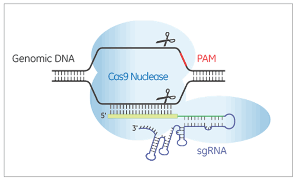
The Dharmacon Edit-R algorithm has been developed and empirically validated to prioritise guides that generate functional protein knockout. Each predesigned guide RNA is scored for two key factors:
- Functionality: the likelihood of producing protein-level knockout
- Specificity: the predicted risk of off-target editing
This gives researchers a practical way to balance knockout performance and specificity depending on their cell model, target gene and downstream assay.
Guide RNA formats to match different CRISPR workflows
The Edit-R platform supports several guide RNA formats, allowing researchers to choose a workflow that suits their experimental setup rather than forcing every project into the same delivery strategy.
Synthetic crRNA system
The synthetic crRNA workflow uses three core components:
- A source of Cas9 nuclease
- A gene-specific Edit-R crRNA
- The Edit-R tracrRNA scaffold
The tracrRNA complexes with the gene-specific crRNA to direct Cas9 to the target site. This cloning-free approach helps researchers move from design to editing quickly, without needing vector construction or sequencing confirmation before starting an experiment.
This format is well suited to researchers who want a flexible, modular workflow and the ability to pair synthetic guide RNAs with different Cas9 delivery options, including purified protein, mRNA or lentiviral Cas9 expression.
Synthetic sgRNA
For researchers who prefer a single-guide format, Edit-R synthetic sgRNA combines the crRNA and tracrRNA sequences into one molecule. This simplifies handling and reduces the number of components in the workflow while retaining compatibility with DNA-free Cas9 delivery strategies.
Synthetic sgRNA can be a useful option when workflow simplicity, transfection consistency and rapid experimental setup are priorities.
Lentiviral sgRNA vectors
For stable, long-term expression, particularly in difficult-to-transfect cells or for follow-up validation after pooled screening, Edit-R lentiviral sgRNA vectors provide a more durable delivery approach.
The Edit-R lentiviral sgRNA vector expresses the gene-specific crRNA and tracrRNA as a chimeric single guide RNA under the control of a human U6 promoter. A puromycin resistance marker, driven from the mouse CMV promoter, is co-expressed in the same vector. This allows researchers to select cells carrying the integrated sgRNA construct.
This format is especially useful for:
- Difficult-to-transfect cell types
- Stable knockout studies
- Follow-up validation from pooled CRISPR screens
- Experiments requiring longer-term guide RNA expression
- Workflows where antibiotic selection is useful for enriching edited populations
All-in-one lentiviral sgRNA
The all-in-one lentiviral sgRNA format takes the stable expression workflow one step further by combining Cas9 and sgRNA expression in a single vector. This reduces the number of transduction steps required and can simplify workflow optimisation, particularly where introducing separate Cas9 and guide RNA components would add time or variability.
For researchers working in challenging cell models, the all-in-one format may provide a more streamlined route to stable CRISPR knockout experiments.
Human guide RNAs realigned to the latest RefSeq database
In late 2025, all predesigned human Edit-R synthetic and lentiviral guide RNAs were realigned against the latest NCBI RefSeq database.
This update recalculates functionality and specificity scores using the current genomic reference. Any designs that no longer met the required scoring threshold were replaced with higher-scoring alternatives.
For most genes, previously used guides remain unchanged. Where a guide has been retired, the replacement reflects a stronger predicted balance of functionality and specificity based on updated genome annotations.
Researchers who have previously ordered Edit-R guide RNAs can check specific catalogue numbers through the Horizon Discovery website. This is useful if you are repeating an earlier experiment, comparing results across projects or planning follow-up work based on a previously ordered guide.
New Cas9 Protein Hybrid NLS for DNA-free RNP editing
The new Edit-R Cas9 Protein Hybrid NLS is a purified Cas9 nuclease protein featuring an enhanced hybrid nuclear localisation signal composition. It is designed to improve nuclear delivery compared with traditional single-NLS formats.
The protein is optimised for co-electroporation or co-transfection with Edit-R synthetic guide RNAs to form ribonucleoprotein complexes, commonly referred to as RNPs. This enables a completely DNA-free CRISPR editing workflow.
DNA-free RNP editing can offer several practical advantages:
- No plasmid DNA is introduced into the system
- The risk of plasmid integration is avoided
- Promoter compatibility issues are removed
- Cas9 exposure is transient, helping to reduce off-target exposure
- The workflow can be useful for sensitive or clinically relevant cell models
Functional knockout has been demonstrated in primary human CD4+ T cells by nucleofection, and in U2OS cells using both nucleofection and lipid transfection.
The Edit-R Cas9 Protein Hybrid NLS is available in sizes from 50 µg to 5 × 500 µg, with bulk quantities available on request.
Where CRISPR knockout workflows can be applied
For molecular biology researchers, the value of these updates is not that CRISPR is new. It is that more current guide design and more flexible Cas9 delivery options can help improve the reliability of familiar workflows.
Edit-R reagents can support applications such as:
- Functional genomics studies
- Disease modelling
- Pathway analysis
- Target identification and validation
- Pooled screen follow-up
- Primary cell editing
- Cell and gene therapy research workflows
By choosing the appropriate guide RNA format and Cas9 delivery strategy, researchers can better tailor CRISPR knockout workflows to their cell type, experimental timeline and downstream readout.
Edit-R performance guarantee
Every predesigned Edit-R synthetic crRNA, sgRNA, lentiviral sgRNA and all-in-one lentiviral sgRNA is backed by an editing guarantee.
If a predesigned guide does not edit the target site when used as recommended, a replacement guide of the same format and quantity will be provided at no cost.
Key takeaways
The latest Dharmacon Edit-R updates provide researchers with a more current and flexible platform for CRISPR knockout experiments.
- Edit-R guide RNAs are designed for functional protein knockout, not just indel generation
- Predesigned human guide RNAs were realigned to the latest RefSeq database in late 2025
- Multiple guide RNA formats support synthetic, lentiviral and all-in-one CRISPR workflows
- Lentiviral formats provide stable expression options for difficult-to-transfect cells and longer-term studies
- The new Cas9 Protein Hybrid NLS supports DNA-free RNP editing with enhanced nuclear delivery
- Edit-R predesigned guide RNAs are backed by an editing guarantee
Explore Dharmacon Edit-R CRISPR solutions
Millennium Science supplies Revvity’s Dharmacon Edit-R CRISPR platform across Australia and New Zealand, supporting researchers with genome editing reagents, workflow selection and technical guidance.
Interested in learning more?
- Request pricing for Edit-R CRISPR reagents
- Discuss the best workflow for your cell model
- Explore DNA-free RNP editing strategies
- Speak with a CRISPR product specialist
On this page
- What’s New in the Dharmacon™ Edit-R™ CRISPR Platform
- Designed for functional protein knockout
- Guide RNA formats to match different CRISPR workflows
- Human guide RNAs realigned to the latest RefSeq database
- New Cas9 Protein Hybrid NLS for DNA-free RNP editing
- Where CRISPR knockout workflows can be applied
- Edit-R performance guarantee
- Key takeaways
- Explore Dharmacon Edit-R CRISPR solutions
Agilent Liquid Handling Instruments: A Complete Guide to Readers, Washers & Automation
Agilent Liquid Handling & Microplate Instrument Portfolio: Choosing the Right Workflow Solution for Your Lab
Modern life science laboratories are under constant pressure to generate reliable data faster, improve reproducibility, and streamline increasingly complex workflows. From ELISA automation and nucleic acid quantification to high-throughput screening and live-cell analysis, selecting the right liquid handling and detection platform can significantly improve laboratory efficiency.
Agilent’s liquid handling and microplate instrumentation portfolio combines flexible automation, multimode detection, plate washing, dispensing, and workflow integration technologies designed to support research laboratories ranging from routine assay environments through to advanced high-throughput screening facilities.
Why Agilent Liquid Handling Systems?
Agilent offers a broad range of workflow solutions that help laboratories:
- Reduce manual pipetting and repetitive handling
- Improve assay consistency and reproducibility
- Scale from low-throughput to automated high-throughput workflows
- Support applications including ELISA, cell-based assays, nucleic acid quantification, microbial kinetics, phenotypic screening, and organoid workflows
- Integrate automation across washing, dispensing, detection, and plate handling
How to Choose the Right Agilent Liquid Handling Solution
The ideal platform depends on your workflow complexity, throughput requirements, and assay types.
| Workflow Need | Recommended Solutions |
| Routine ELISA workflows | 800 TS + 50 TS |
| Nucleic acid quantification | Epoch or Epoch 2 |
| Flexible multimode detection | Synergy LX or Synergy H1 |
| High-throughput screening | Synergy Neo2 + BioStack |
| Automated wash/dispense workflows | EL406 or 406 FX |
| 3D cell culture dispensing | MultiFlo FX |
| Live-cell imaging and phenotypic analysis | Cytation 9 |
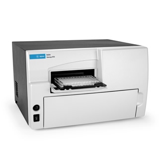
Agilent Absorbance Microplate Readers
Agilent absorbance readers support routine laboratory workflows including ELISAs, protein assays, microbial growth studies, and nucleic acid quantification.
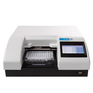
Agilent 800 TS Microplate Reader
The 800 TS is designed for straightforward absorbance workflows and cost-conscious laboratories requiring reliable performance for routine assays.
Key features
- Reads wavelengths from 400-750 nm
- Supports 6- to 384-well plates
- Touchscreen interface with USB export
- Optional temperature control and shaking
- Gen5 software integration
Best-fit applications: ELISAs, Protein assays, Enzyme kinetics, Basic cell-based assays
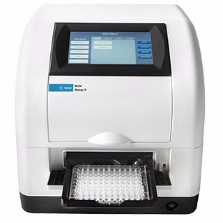
Agilent Epoch Microplate Spectrophotometer
The Epoch combines monochromator-based UV-Vis detection with flexible wavelength selection for nucleic acid and protein quantification workflows.
Key features
- UV-Vis range from 200-999 nm
- Compatible with Take3 micro-volume plates
- Temperature control to 65 °C
- Supports 6- to 384-well plates
Best-fit applications: DNA and RNA quantification, 260/280 and 260/230 purity measurements, Cytotoxicity assays, Cell proliferation studies, Enzyme kinetics
Agilent Epoch 2 Microplate Spectrophotometer
The Epoch 2 expands absorbance capabilities with stand-alone touchscreen operation and full-spectrum scanning functionality.
Ideal for: Spectral scanning, Microbial growth kinetics, Nucleic acid purity analysis, Stand-alone absorbance workflows
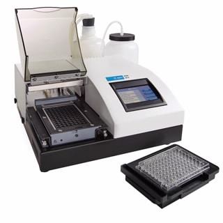
Automated Plate Washers for ELISA & Cell-Based Assay
Reliable washing is critical for reducing background noise and improving assay consistency in ELISA and cell-based workflows.
Agilent 50 TS Washer
The 50 TS provides dependable automated washing for routine assays and pairs naturally with absorbance readers like the 800 TS.
Applications: ELISA washing, Cell-based assays, Vacuum filtration workflows
Agilent 405 LS and 405 TS Washers
These automated plate washers support more advanced workflows with dual-action manifold technology and automation compatibility.
Key features
- Automated washing for 96- and 384-well plates
- Biomagnetic separation support
- Vacuum filtration modules
- Four-buffer switching
- BioStack automation compatibility
Best-fit workflows: ELISA, Microsphere-based assays, Automated cell-based assays
Agilent Washer-Dispenser Systems
Combining washing and dispensing into a single platform can simplify workflows and reduce instrument footprint.
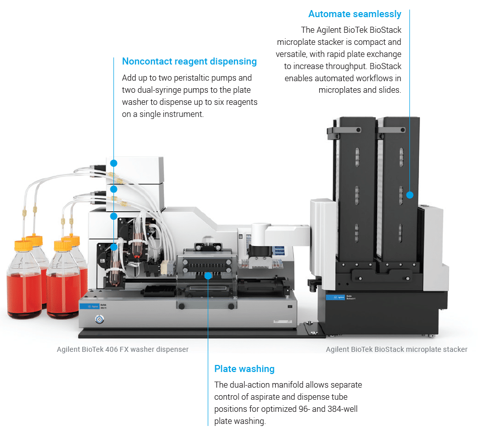
Agilent 406 FX Washer Dispenser
The 406 FX integrates washing and dispensing capabilities for automated multi-step assay workflows.
Highlights
- Washing plus up to six reagent dispensers
- Supports biomagnetic protocols
- Compatible with 96- to 1536-well plates
- Robotic integration support
Common applications: ELISA automation, Cell culture workflows, Bead-based assays
Agilent EL406 Washer Dispenser
The EL406 combines rapid washing and dispensing into a compact automation-ready platform.
Ideal for: High-throughput ELISA workflows, Multiplex assays, Integrated wash and dispense automation
Bulk Reagent Dispensing
Agilent MultiFlo FX Multi-Mode Dispenser
The MultiFlo FX is designed for flexible reagent dispensing across a wide range of assay formats.
Key capabilities
- Dispensing to 6- to 1536-well plates
- Up to four reagents in parallel
- Gentle media exchange options for delicate cultures
Applications: 2D and 3D cell culture, Spheroid and organoid workflows, ELISA preparation, Bead-based assays
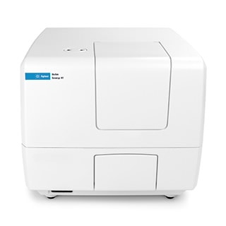
Multimode Readers for Advanced Detection
Agilent multimode readers combine absorbance, fluorescence, and luminescence detection technologies to support a broad range of assay requirements.
Agilent Synergy Neo2
The Synergy Neo2 is designed for demanding high-throughput and multiplex workflows.
Features
- Hybrid detection with filter and monochromator optics
- Laser TRF capability
- Up to four PMTs
- Environmental control with CO₂/O₂ regulation
Applications: High-throughput screening, Multiplex assays, Advanced multimode workflows
Agilent Synergy HTX
The Synergy HTX provides economical multimode detection for routine assay environments.
Detection modes: Absorbance, Fluorescence, Luminescence, Alpha assays
Agilent Synergy LX
The Synergy LX is designed for straightforward multimode detection with an intuitive touchscreen interface.
Ideal applications: Nucleic acid quantification, ELISA, BCA and Bradford assays, Cell viability workflows
Agilent Synergy H1
The modular Synergy H1 platform enables laboratories to scale capabilities as assay requirements evolve.
Supports: Absorbance, Fluorescence, Luminescence, AlphaScreen, TRF workflows
Imaging & High-Content Analysis Workflows
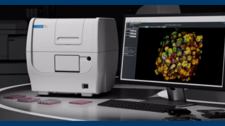
Agilent Cytation 9
The Cytation 9 combines automated imaging with multimode detection in a single platform.
Key capabilities
- Imaging up to 60× magnification
- Fluorescence, luminescence, and UV-Vis detection
- Live-cell environmental control to 65 °C
- BioStack automation compatibility
Common applications: Live-cell imaging, Phenotypic screening, Quantitative cell analysis
High-Throughput Laboratory Automation
Agilent BioStack
The BioStack automates plate loading and unloading for walk-away workflow automation.
Benefits
- Fast plate exchange
- Delidding and re-lidding support
- Compatible with 6- to 1536-well plates
- Enables unattended high-throughput processing
Supporting Modern Research Workflows
Agilent’s liquid handling and microplate instrumentation portfolio supports a wide spectrum of laboratory applications, from routine absorbance assays through to advanced automation and high-content workflows. With scalable solutions for washing, dispensing, detection, imaging, and plate automation, laboratories can build flexible workflows that improve reproducibility, efficiency, and throughput.
To discuss the best Agilent liquid handling workflow for your laboratory, contact the team at Millennium Science.
On this page
- Agilent Liquid Handling & Microplate Instrument Portfolio: Choosing the Right Workflow Solution for Your Lab
- Why Agilent Liquid Handling Systems?
- How to Choose the Right Agilent Liquid Handling Solution
- Agilent Absorbance Microplate Readers
- Automated Plate Washers for ELISA & Cell-Based Assay
- Agilent Washer-Dispenser Systems
- Bulk Reagent Dispensing
- Multimode Readers for Advanced Detection
- Imaging & High-Content Analysis Workflows
- High-Throughput Laboratory Automation
- Supporting Modern Research Workflows
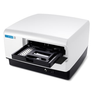
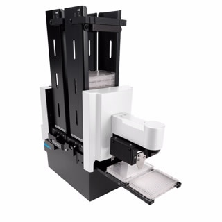
Seeing More, Doing More: Agilent’s BioTek Imaging Portfolio in Action!
A Joe Blogs post by Joe Roberts, PhD
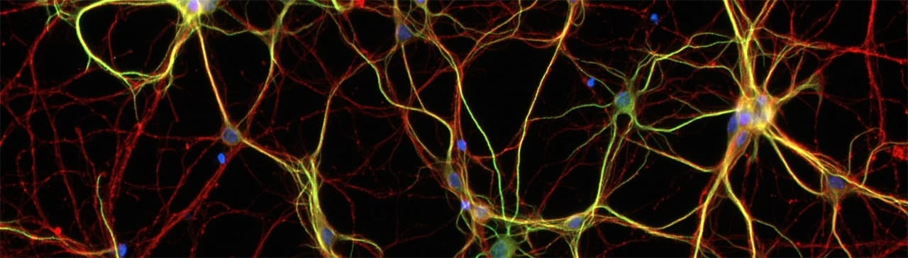
Modern life science research increasingly demands more information from fewer samples, delivered faster and with greater confidence. Whether you’re studying immune cell function, complex 3D models, or subtle phenotypic changes, imaging is central to generating meaningful insights. Agilent’s BioTek imaging portfolio is designed to meet this challenge, combining high-quality microscopy, flexible multimode detection, and live-cell capability into integrated platforms that scale with your research needs.
Cytation and Lionheart: Imaging Platforms That Scale With Your Research
The Agilent BioTek Cytation™ range spans entry-level widefield imaging through to advanced confocal workflows, combining automated microscopy with multimode microplate reading in a single, integrated platform. Designed to scale with experimental complexity, Cytation systems support routine plate-based assays, high-content imaging, and live-cell experiments with environmental control, making them suitable for laboratories of all sizes and budgets.
For labs requiring dedicated automated microscopy, the Agilent BioTek Lionheart™ FX and LX microscopes deliver high-performance imaging in a compact footprint. These systems support a wide range of objectives and imaging modalities, with robust autofocus, flexible incubation options, and automation-ready workflows. Lionheart platforms are particularly well suited to phenotypic profiling, kinetic assays, and live-cell imaging where precision and repeatability are essential.
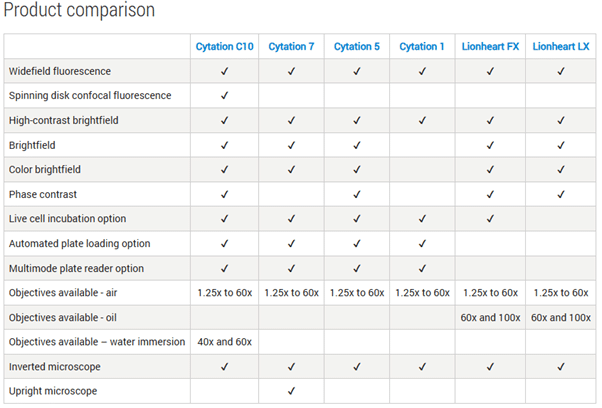
A detailed product comparison table below highlights the specific capabilities of each Cytation and Lionheart model.
Why Agilent BioTek Imaging Stands Out
Across the portfolio, Agilent imaging platforms share several key advantages:
- Support for 6- to 1536-well plates, slides, dishes, and flasks.
- Compatibility with automation solutions like BioStack™, BioSpa™, and BenchCel™.
- Live-cell imaging with CO₂/O₂ control, shaking, and temperature incubation.
- Advanced imaging capabilities: Z-stacking, Z-projection, montage stitching, and time-lapse videos.
- Hybrid optics for sensitive and specific fluorescence detection.
Applications include wound healing assays, cell migration and invasion studies, immunofluorescence, neurite outgrowth analysis, 3D cell models, and ADME/Tox experiments. By unifying imaging and detection in a single platform, Agilent BioTek systems help labs move beyond single-endpoint measurements to richer, biologically meaningful data.
Joe’s Takeaway
Whether you’re starting with simple plate-based assays or pushing the boundaries of confocal or automated microscopy, the Agilent BioTek imaging portfolio gives you the flexibility, performance, and scalability to see more and do more.
If you’re interested in a demo, contact us today!
Until next time… happy experimenting!

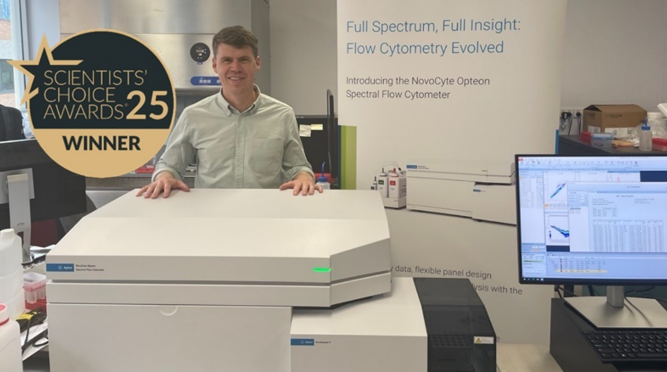
Unleashing Spectral Power: Agilent NovoCyte Opteon Arrives in ANZ
A Joe Blogs post by Joe Roberts, PhD
Spectral flow cytometry continues to transform single-cell research – and now, for the first time, laboratories in Australia and New Zealand can experience the Agilent NovoCyte Opteon first hand.
Launched globally at CYTO 2024, the NovoCyte Opteon has already earned international recognition, taking home the Select Science Scientist’s Choice Award for “Best New Drug Discovery & Development Product of 2024.” With up to five lasers, 73 detectors, and cutting-edge optical engineering, this system represents the next generation of high-dimensional cytometry – and it’s now available for demo in ANZ.
Spectral acquisition, redefined
Unlike conventional cytometry, the NovoCyte Opteon captures the full emission spectra of each fluorochrome across all lasers. This approach allows for greater panel flexibility, more accurate unmixing, and cleaner resolution of overlapping signals – ideal for complex immunophenotyping studies.
Spectral Flow Cytometry vs Conventional Flow: What’s the Difference?
Traditional flow cytometry detects fluorescence using individual optical filters for each fluorochrome, limiting panel size and often causing signal overlap.
Spectral flow cytometry – like the Agilent NovoCyte Opteon – captures the entire emission spectrum from every fluorochrome across all lasers.
This enables:
-
Greater panel flexibility with more markers in a single run
-
Improved accuracy through spectral unmixing of overlapping signals
-
Cleaner data by accounting for autofluorescence as a separate spectral component
The result is a more powerful, precise, and reproducible analysis – ideal for complex immunophenotyping and high-dimensional research.
Configurability and Optics
Researchers can choose 3-, 4-, or 5-laser configurations, with the flagship system spanning UV, Violet, Blue, Yellow/Green, and Red lasers – and up to 73 detectors. Agilent’s proprietary optics and electronics maximise sensitivity and spectral separation, ensuring high-quality data.
Dynamic Range, Small-Particle Detection, and Autofluorescence Handling
It boasts a wide dynamic range both for fluorescence and scatter (size) detection, reducing the need for frequent detector adjustments.
Dual-laser small particle detection (using 405 nm and 488 nm SSC) enables detection of particles down to ~80 nm without needing separate adjustments between cell and particle modes.
Also, the instrument supports autofluorescence subtraction (i.e. treating autofluorescence as a spectral component), which helps resolve dim populations more clearly.
Reliability and Stability Built In
To maintain performance in variable lab environments, the NovoCyte Opteon integrates on-board temperature control, fluidics monitoring, electronics sensor circuits, and real-time instrument status feedback.
Automation and Throughput
It’s compatible with the NovoSampler S, accepting 40-tube racks and microplates (384/96/48/24), and is ready for robotic automation. Calibration is automated, with templates for labware types saved for reproducibility. Carryover is minimal (< 0.1 %) via rinse cycles.
Software and Workflows
Agilent’s NovoExpress (Opteon) software version 2.0+ underpins the acquisition, unmixing, analysis, and reporting workflow. They’ve enhanced the user interface with an “unmixing” tab, streamlining spectral unmixing steps. The software supports both real-time acquisition and downstream “offline” analyses.
Joe’s Takeaway
The arrival of the Agilent NovoCyte Opteon in the ANZ region marks a real milestone for spectral flow cytometry. Local researchers now have access to one of the most advanced, award-winning platforms available – combining powerful optics, automation readiness, and Agilent’s renowned reliability, all supported locally by Millennium Science.
If you’re interested in demoing the award-winning NovoCyte Opteon Spectral Flow Cytometer, we’d love to hear from you. Reach out to arrange a hands-on session and see what spectral flow can really do for your research.
And if you’re attending CYTO-Connect (Perth, November 27-29, 2025), come and say hello – we’ll be there showcasing the Opteon and chatting all things spectral!
If you’re interested in a demo of the Opteon, contact us today!
Until next time… happy experimenting!
On this page
- A Joe Blogs post by Joe Roberts, PhD
- Spectral acquisition, redefined
- Spectral Flow Cytometry vs Conventional Flow: What’s the Difference?
- Configurability and Optics
- Dynamic Range, Small-Particle Detection, and Autofluorescence Handling
- Reliability and Stability Built In
- Automation and Throughput
- Software and Workflows
- Joe’s Takeaway
Unlocking the Power of Spatial Biology with the Right Antibody Choices
Secondary Antibody Selection for Spatial Biology
Spatial biology is transforming the way we understand biological systems. By integrating spatial information into research, it provides a holistic view of how cells and molecules interact within their native environment. This approach sheds light on the complex interplay between cellular and molecular components, offering deeper insights into the function and behaviour of living organisms.
To achieve reliable and reproducible results in spatial biology, careful antibody selection is essential. Here are some key considerations:
Consider Host and Target Species
Secondary antibody selection begins with species. The host of your secondary must differ from both the tissue species and the host of the primary antibody. For example, when using a rabbit primary antibody on human tissue, a goat anti-rabbit secondary is preferred. This reduces background interference and ensures the signal reflects true binding rather than cross-reactivity..
Match Secondary to Primary Class
Not all primary antibodies are the same. Polyclonal IgGs require anti-IgG secondaries, while monoclonal IgMs need anti-IgM secondaries. For monoclonal IgG subclasses (such as IgG1), it is best practice to use subclass-specific secondaries (anti-IgG1) for maximum accuracy. This level of matching safeguards against nonspecific binding and strengthens reproducibility.
Affinity-Purified: Cut the Noise
Affinity purification ensures secondaries recognise their targets with high specificity. By removing unwanted immunoglobulins, affinity-purified antibodies deliver clearer signals and consistent results – especially when detecting low-abundance proteins. The result: less noise, less background, and greater confidence in your data.
Cross-Adsorbed for Multiplexing
Spatial biology thrives on multiplexing, but multiple species and fluorophores introduce complexity. Cross-adsorbed antibodies are refined to remove cross-reactive components, lowering background and minimising false positives. This makes them ideal for multi-label experiments where precision is paramount.
Choose the Right Fluorophores
Signal clarity depends on the brightness and stability of your fluorophores. Rockland offers a wide range of conjugates, including DyLight™, Cy™, and FITC dyes. These high-performance labels are particularly powerful for detecting low-expression targets, ensuring that no signal is missed in complex tissue environments.
A Case in Point: Neural Crest Imaging
In one example, Rockland’s DyLight™ 649-conjugated goat anti-rat secondary antibodies were used to visualise neural crest-derived cells infiltrating the brain region of a mouse embryo. The result was a sharp, specific signal that allowed researchers to trace cell migration and interactions in detail. This illustrates the impact that well-chosen secondary antibodies can have on spatial imaging outcomes.
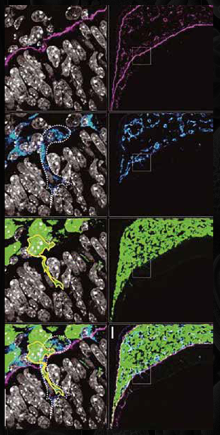
Conclusion
When it comes to spatial biology, success is in the details. Rockland’s secondary antibodies – affinity-purified, cross-adsorbed, and conjugated to high-performance fluorophores – provide the specificity and sensitivity needed for reproducible results.
Introducing Atlas™: The Next Era of Cell Imaging
Better data. Faster breakthroughs. Confident choices.
LICORbio’s new Atlas™ Imager is redefining cell imaging for life science researchers across Australia and New Zealand. Built for both 2D and 3D assays, Atlas combines speed, scale, and clarity in a single, easy-to-use platform – empowering scientists to accelerate discoveries in drug development, translational research, and beyond.
Smarter, Faster, Scalable Imaging
Unlike traditional systems that require manual stitching or time-consuming illumination corrections, Atlas is engineered to deliver whole-plate imaging in less than one minute. Its patented line-scanning optical system minimises background fluorescence, boosts sensitivity, and enhances multiplexing capabilities – critical for high-throughput screening and complex cell-based assays.
Key advantages of the LICORbio Atlas
- High throughput – Acquire entire well plates rapidly or zoom into single wells with up to 5µm resolution.
- Broad applications – Capture cell viability, luminescence, and multiplex fluorescence data in one instrument.
- Deep 3D imaging – Confidently image spheroids, organoids, organ-on-a-chip models, and other microphysiological systems.
- Ease of use – Streamlined workflows reduce manual steps and consolidate multiple instruments into one solution.
- Cost-effective – Higher-content imaging without the high price tag of competitive systems.
Designed for Today’s Translational Research
From monitoring cell culture to high-resolution plate and well scanning, Atlas provides reliable, reproducible, and quantitative results with minimal hands-on time. With over 30 imaging channels spanning UV to near-infrared, researchers gain unmatched flexibility for diverse applications – from drug screening to advanced tissue modelling.
By eliminating inefficiencies and post-processing steps, Atlas lets you focus on what matters most: generating insights that move science forward.
Why Atlas Stands Out
- Whole-plate scans in under one minute
- Z-stack multiplex fluorescence for 3D studies
- Built-in luminescent imager
- Over 30 imaging channels with six lasers plus RGB LED
- End-to-end solution for cell-based assays
Accelerate Your Next Discovery
Whether you’re developing new therapeutics, studying disease models, or advancing organoid research, Atlas is the balanced solution for speed, sensitivity, and scalability in cell imaging.
The Reagent Insider: Advancing Alzheimer’s research
Tips, insights and updates
As the leading cause of dementia, Alzheimer’s disease impacts over 50 million people worldwide. In Australia, dementia has recently overtaken heart disease as the leading cause of death, particularly among women. While current treatments focus on symptoms, new research aims to target the underlying disease from clearing amyloid-β, inhibiting tau aggregation, and modulating neuroinflammation to understanding the gut-brain axis and early metabolic changes in AD.
Hot Product
StressMarq’s high-quality tau, amyloid-β, and alpha-synuclein proteins support research into the mechanisms underlying Alzheimer’s disease. Contact us for details.
![]()
Exploring the Gut-Brain Link in Alzheimer’s
Emerging studies show gut health influences cognitive function, with a balanced microbiome and Mediterranean diet potentially lowering dementia risk. Using Norgen Biotek’s Stool Nucleic Acid Collection and Preservation Systems, researchers are uncovering novel approaches to tackle cognitive decline.
Connect with Narisa Dawar to learn more about Norgen Biotek.
Lab Favourite
Silence Alzheimer’s-related genes with siRNA.
Targeting tau reveals its role in microtubule stability and neurofibrillary tangles. Check out Revvity’s Dharmacon™ siRNA solutions.
MedChemExpress Neurodegenerative Disease-related Compound Library
This library of 2,960 compounds targets amyloid-β, dopamine receptors, COMT, LRRK2, 5-HT receptors, and more! Perfect for high-throughput, high-content screening of neurodegenerative mechanisms and potential treatments for Alzheimer’s, Parkinson’s, and MND.
Curious about MCE’s compound libraries? Christine Goy is just an email away!
Protocol Tip
Stay on track in Alzheimer’s research. Use InvivoGen’s Mycoplasma Strip to detect contamination early to ensure reliable neuronal and glial culture results. Email Christine Goy for details.
Metabolomics and the Brain
Understanding early metabolic changes in Alzheimer’s disease may identify new therapeutic targets and predictive biomarkers. Metabolomics can detect these subtle biological shifts before clinical symptoms appear, supporting the development of early interventions and treatments that may improve outcomes for people with AD.

Advance Your Infectious Disease Research with ACROBiosystems
Power Your Research: Proteins, Antibodies & More Await!
As we step into the new year, Millennium Science is excited to partner with you in advancing your research! Whether you’re embarking on groundbreaking projects or tackling routine experiments, our exclusive selection of ACROBiosystems reagents for infectious disease research is designed to support every stage of your work. With special offers on premium products, we are committed to providing you with the tools needed to achieve precise, reliable results in virology and immunology studies.
Why Choose ACROBiosystems Reagents for Infectious Disease Research?
ACROBiosystems is a trusted global leader in the development of high-quality recombinant proteins, antigens, antibodies, and assay kits for infectious disease research. Their reagents are widely used in vaccine development, antibody screening, diagnostic assay development, and therapeutic drug discovery. With a comprehensive portfolio spanning immunology, oncology, and virology, ACROBiosystems ensures reproducibility and accuracy in scientific experiments.
Key Benefits of ACROBiosystems Reagents:
- High Purity & Bioactivity – Ensuring reliable and consistent experimental results.
- Extensive Product Portfolio – Covering a wide range of infectious disease targets, including SARS-CoV-2, influenza, RSV, HPV, and more.
- Custom Solutions Available – Tailored reagents to meet specific research needs.
- Validated for Diagnostic and Therapeutic Applications – Trusted by leading biotech and pharmaceutical companies.
Millennium Science is a proud distributor of ACROBiosystems, offering exclusive access to their latest products and technical expertise.
The Global Impact of Infectious Diseases
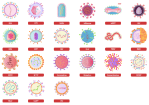
Infectious diseases, caused by viruses, bacteria, fungi, and parasites, continue to pose significant global health risks. These diseases can spread through direct human contact, contaminated water, airborne transmission, and insect vectors. Some of the most pressing infectious diseases include:
- Respiratory Viruses – Influenza, respiratory syncytial virus (RSV), SARS-CoV-2
- Zoonotic Diseases – Rabies, Hendra virus, Nipah virus
- Oncogenic Viruses – Human papillomavirus (HPV), Epstein-Barr virus (EBV)
- Tropical & Vector-Borne Diseases – Malaria, dengue fever, chikungunya
With the ongoing threat of pandemics and emerging viral infections, researchers worldwide are focused on vaccine development, antiviral drug discovery, and improved diagnostic methods.
Addressing Viruses of Concern with Advanced Reagents
Since late 2019, the SARS-CoV-2 pandemic has highlighted the importance of robust virology research and vaccine development. Historically, diseases like smallpox, the plague, and the Spanish flu have had profound impacts on human society. Today, viruses such as influenza, rabies, RSV, and HPV continue to threaten global health.
To combat these challenges, scientists rely on high-quality recombinant proteins, neutralising antibodies, and assay development tools to accelerate vaccine research and therapeutic discoveries.
Our Commitment to Advancing Infectious Disease Research
At Millennium Science, we are dedicated to supporting virologists, immunologists, and infectious disease researchers with cutting-edge reagents that drive progress. Our partnership with ACROBiosystems enables us to provide:
- Top-tier reagents for virus research, vaccine development, and drug discovery.
- Access to the latest recombinant proteins and antigens.
- Ongoing technical support and consultation for product selection and protocol optimisation.
Explore Our Reagents for Infectious Disease Research
Click below to search our extensive catalogue of ACROBiosystems reagents for your infectious disease research, and discover how Millennium Science can support your next breakthrough!
On this page
- Power Your Research: Proteins, Antibodies & More Await!
- Why Choose ACROBiosystems Reagents for Infectious Disease Research?
- The Global Impact of Infectious Diseases
- Addressing Viruses of Concern with Advanced Reagents
- Our Commitment to Advancing Infectious Disease Research
- Explore Our Reagents for Infectious Disease Research
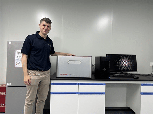
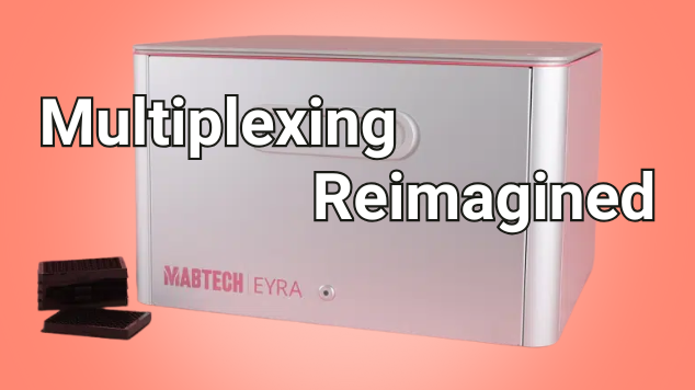
Meet EYRA: Multiplexing Reimagined
A Joe Blogs post by Joe Roberts, PhD
When it comes to multiplex protein analysis, researchers need accuracy, speed, and simplicity. Traditional flow-based systems have long been the standard, but they bring challenges: sheath fluids, blocked probes, and constant maintenance. Enter the Mabtech EYRA™ – a fluidics-free multiplex immunoassay platform that reimagines how scientists generate cytokine and biomarker data. With confocal imaging, RAWsphere analysis, and compatibility with EYRAplex bead kits, EYRA makes multiplexing faster, simpler, and more reliable.
Multiplex Without Compromise
With EYRAplex magnetic bead assays, EYRA can quantify more than 30 analytes from a single sample – whether that’s serum, plasma, or cell culture supernatant. This means less sample consumption, fewer runs, and richer datasets for every experiment.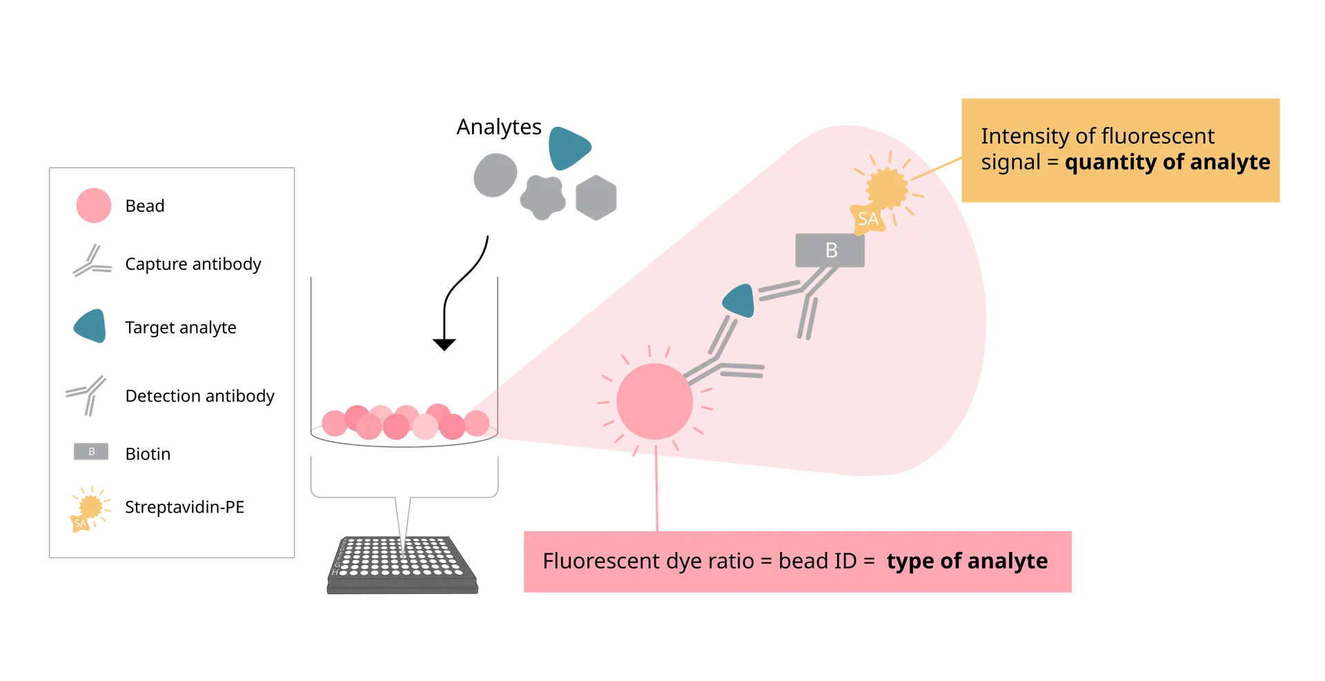
At the core of EYRA’s precision is the RAWsphere image analysis algorithm. It identifies each bead, links it to the correct analyte, and quantifies the PE signal with high resolution. Whether you’re measuring six cytokines or a full 30+ panel, RAWsphere ensures your multiplex data is accurate and reproducible.
Watch video to see how EYRA works
Fluidics-Free Technology
EYRA is built around a completely flow-free design. No sheath fluid, priming, waste handling or blocked probes.
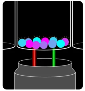
Instead of pushing samples through fluidics, EYRA uses confocal microscopy to image settled magnetic beads directly in the wells of a 96-well plate. Each bead carries a unique fluorescent dye signature for identification, while analyte-bound PE-labelled antibodies provide the quantitative signal.
The result? You insert your plate, select your assay in the intuitive Mabtech Opal™ software, and hit read. Fifteen minutes later, you have fully processed data – with your samples never leaving the wells.
From Plate to Excel - Fast
Opal™ software comes preloaded with templates for every EYRAplex kit. Standards, plate layouts, and gating are handled automatically – so you spend less time setting up and more time on results.
Once the plate read is complete, results are exported directly into Excel, with options for bulk export if you’re working on large studies or multiple plates. No manual reformatting. No data wrangling headaches.
The Maintenance-Free Mindset
Because EYRA is fluidics-free, there’s no daily calibration or cleaning. No flushing, no wasted consumables, and no time lost to instrument downtime. It’s genuinely a plug-and-play experience – switch it on, run your plate, and walk away with your data.
Joe’s Takeaway
The Mabtech EYRA isn’t just another multiplex platform – it’s multiplexing reimagined. By removing fluidics and harnessing high-resolution confocal imaging, EYRA delivers:
- High-plex capacity: 30+ analytes per well
- Fast turnaround: ~15 minutes per plate
- No maintenance: no daily cleaning or calibration
- Accurate, reproducible results: powered by RAWsphere analysis
For labs looking to streamline their workflow without sacrificing data quality, EYRA offers a fresh, frustration-free alternative to traditional flow-based systems.
Until next time… happy experimenting!
Joe Roberts, PhD
Product Manager
Millennium Science
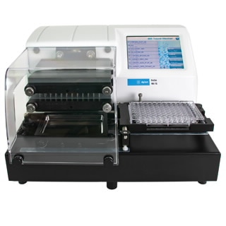
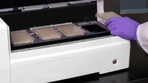
Streamline Microbial Growth Studies with the Agilent BioTek LogPhase 600
Overview
The Agilent BioTek LogPhase™ 600 Microbiology Reader is a high-performance solution for labs studying microbial growth and metabolism. Purpose-built to support real-time, kinetic analysis of bacteria and yeast cultures, the LogPhase 600 enables the simultaneous monitoring of microbial growth curves across up to four standard 96-well microplates – boosting throughput and consistency for microbiology applications.
Precision Temperature Control and Shaking for Reliable Growth Conditions
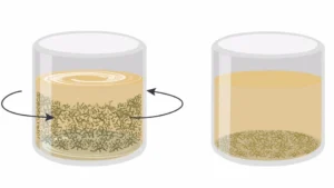
Designed specifically for microbial assays, the LogPhase 600 features robust shaking and sensor-driven temperature control, which are essential for optimal bacterial and yeast cell growth. The integrated orbital shaking mechanism keeps microbial cultures in suspension, promoting uniform growth across all wells.
Advanced temperature regulation ensures uniform heating throughout the instrument, eliminating edge effects and minimising evaporation. A carefully engineered top-to-bottom temperature gradient prevents condensation on sealed plates – reducing light scatter and ensuring accurate optical density (OD) measurements.
Consistent and Reproducible Microbial Growth Curves
Whether you’re performing bacterial growth curve analysis or extended kinetic microbial studies, the LogPhase 600 provides reproducible results across replicates. Its stable environment makes it ideal for long-term microbial monitoring, delivering high-quality data for even the most demanding experimental conditions.
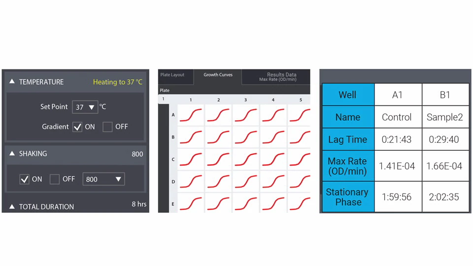
Intuitive Software for Simplified Microbiology Analysis
The LogPhase 600 is controlled via a user-friendly app that simplifies data acquisition and analysis across multiple microplates. Designed with microbiologists in mind, the software allows users to:
-
Start using the instrument with minimal training
-
View multiplate data simultaneously
-
Automatically calculate key growth parameters like Lag time, Maximum growth rate (OD/min) and Time to stationary phase
This makes it easy to integrate the instrument into both academic and industrial lab workflows.
Versatile Applications for High-Throughput Microbiology
With its four-plate capacity, the LogPhase 600 delivers higher throughput than single-plate readers – ideal for labs running large-scale experiments. Common research areas include:
-
Bacterial and yeast growth assays
-
Algal growth studies
-
Antimicrobial resistance research
-
Biofuel production monitoring
-
Food and beverage microbiology testing
Why Choose the Agilent BioTek LogPhase 600?
For researchers seeking a dedicated microbiology plate reader that delivers accuracy, scalability, and ease of use, the LogPhase 600 offers an unmatched combination of throughput, data quality, and application-specific performance. Whether you’re monitoring bacterial growth under various conditions or conducting resistance assays, the LogPhase 600 ensures your data is consistent and actionable.
Contact Us
If you have a question or would like pricing for a LogPhase 600 simply email customerservice@mscience.com.au.
On this page
- Overview
- Precision Temperature Control and Shaking for Reliable Growth Conditions
- Consistent and Reproducible Microbial Growth Curves
- Intuitive Software for Simplified Microbiology Analysis
- Versatile Applications for High-Throughput Microbiology
- Why Choose the Agilent BioTek LogPhase 600?
- Contact Us
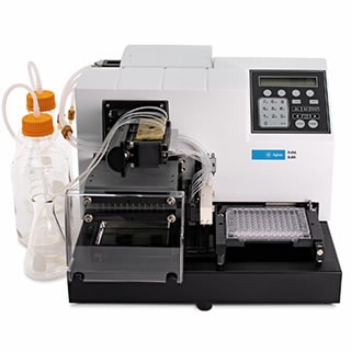
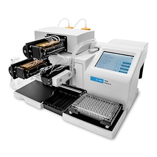
Revolutionising RNA purification with Automation
Accelerating molecular workflows has never been easier
Norgen Biotek’s Magnetic Bead-Based Total RNA Purification Kit is now fully automated, making high-throughput RNA extraction faster, more reliable, and easier to scale.
Designed to isolate all RNA sizes – including microRNA and small RNA – with exceptional purity and reproducibility, this kit delivers consistent performance across a wide range of sample types. Whether you’re working with tissues, cells, bacteria, viruses, bodily fluids, plants or fungi, Norgen Biotek’s advanced RNA purification technology ensures high-yield results without compromising quality.
Save Time with Automated RNA Purification
Manual RNA extraction can be labour-intensive and prone to variability. Norgen Biotek’s magnetic bead-based kit streamlines the process, reducing hands-on time to just 15 minutes, while enhancing consistency across runs.
Built for Lab Automation Platforms
This kit is fully compatible with leading lab automation platforms, including:
-
Opentrons Flex – a modular and cost-effective automation system designed to scale with your lab.
-
Tecan
-
Thermo Fisher KingFisher
-
Hamilton
-
IsoPure
Seamless integration allows labs to boost throughput, minimise human error, and free up valuable time for high-value analysis and discovery.
Why Choose Norgen Biotek RNA Kits?
✅ Compatible with a broad range of sample types
✅ High-quality RNA including microRNA and small RNA
✅ Automation-ready for reliable scalability
✅ Reduced hands-on time and increased reproducibility
Interested in upgrading your RNA workflows?
Explore how Norgen Biotek and Opentrons can modernise your lab’s RNA extraction today or contact Narisa Dawar our Norgen Biotek Product Manager – ndawar@mscience.com.au.
MedChemExpress Anti-Cancer Compound Library
MedChemExpress (MCE) is a supplier of high-quality chemicals, biochemicals and compound libraries, supporting the global scientific research community. MCE’s extensive product portfolio includes a range of screening libraries designed to accelerate drug discovery and development across multiple therapeutic areas. Among these, the Anti-Cancer Compound Library is a powerful tool for researchers seeking targeted solutions and novel cancer therapies.

The library offers over 8,000 compounds with activity against both solid and haematologic tumours, making it suitable for high-throughput screening (HTS) and high-content screening (HCS).
This collection spans key cancer-related pathways such as apoptosis, cell cycle regulation and signal transduction. It also covers diverse signalling pathways – including kinase, GPCR and epigenetics – providing researchers with a comprehensive platform to explore and identify potential anti-cancer agents.
Many of the compounds have undergone rigorous clinical or preclinical evaluations, with some already approved by the FDA, highlighting their therapeutic potential.
The bioactivity and safety profiles of the compounds make the library a useful resource for advancing cancer research. Each compound is also validated for high purity and quality using NMR and LC/MS techniques, which should lead to precise and reproducible results.
A key advantage of MCE’s compound libraries is that users can fully customise a library to meet research needs from the type, quantity and concentrations of compounds and layout, offering the flexibility that researchers need to drive meaningful discoveries in oncology.
Click here to download the MedChemExpress Compounding Libraries brochure or contact Millennium Science at customerservice@mscience.com.au.
Related Blog
MedChemExpress compound libraries for drug discoveryOn this page
Expand Your Lab’s Imaging Capabilities with the LICORbio Odyssey F Imaging System
Accelerated Imaging with Exceptional Sensitivity and Speed
The LICORbio Odyssey F Imaging System delivers high sensitivity and broad dynamic range with just a click. Available in 3- and 10-channel models, this advanced platform scans samples up to twice as fast as previous generations, supporting a wide array of applications beyond standard qualitative Western blotting. Whether you’re imaging proteins, nucleic acids, or other biomolecules, the Odyssey F accelerates your workflow without compromising data quality.
Dual NIR and Visible Lasers for Advanced Multiplex Imaging
Equipped with dual near-infrared (NIR) lasers, the Odyssey F ensures optimal throughput and quantitation for high-quality imaging. In addition, two visible fluorescence lasers unlock powerful multiplexing capabilities, enabling researchers to explore multiple targets simultaneously in plate-based assays, slide imaging, and beyond. This flexibility makes it ideal for researchers aiming to capture more data from a single experiment.
Rapid High-Throughput Imaging with Reproducible Results
Users benefit from rapid scan speeds that reduce total imaging time while improving reproducibility. The Odyssey F is engineered for superior sensitivity and consistent performance, giving researchers increased confidence in their data. Its expanded dynamic range means fewer adjustments and more reliable quantification across a broad concentration spectrum.
Versatile Platform for a Wide Range of Imaging Applications
Designed for versatility, the Odyssey F supports a wide variety of assays and sample types. The expanded focus offset range ensures compatibility with numerous plate formats and sample depths. Whether you’re conducting EMSA/Gel Shift Assays, In-Cell Westerns, Tissue Section Imaging, or Whole Slide Imaging (WSI), the Odyssey F adapts to your lab’s needs, enhancing both productivity and application reach.
See the Odyssey F in action!
Book a demoMeet Aunty from Unchained Labs: The Fastest, Highest Throughput Protein Stability Characterisation Platform on the planet!
About Aunty
Unchained Labs has raised the bar in biologics characterisation with the launch of Aunty – the world’s fastest and most high-throughput platform for protein stability analysis. Tailored for researchers in biotech, pharma, and gene therapy, Aunty delivers unprecedented speed, sensitivity, and flexibility across a wide range of stability parameters – all from a single 96-well quartz plate.

Why Aunty is a Game-Changer in Protein and Viral Vector Stability Testing
🔬 Comprehensive Stability Profiling — In Minutes
Aunty characterises protein melting (Tm, Tonset), aggregation (Tagg, Tsize), colloidal stability (kD, B22, G22), long-term stability, and viral vector genome ejection, reading a full 96-well plate every minute. With only 8 µL sample per well, it saves valuable material while delivering maximum insight.
💡 Three Technologies. One Platform.
- Full-Spectrum Fluorescence – For intrinsic or dye-based detection of thermal unfolding.
- Static Light Scattering (SLS) – Detects early-stage aggregation with exceptional sensitivity.
- Dynamic Light Scattering (DLS) – Monitors particle size, polydispersity, and colloidal behaviour.
These modalities can be run in parallel or independently, offering complete flexibility to tailor your assay to the molecule at hand.
Explore Aunty’s Core Applications
1. Thermal Ramp Analysis
Track unfolding and aggregation simultaneously using fluorescence and SLS. Aunty’s sensitivity enables detection down to 25 µg/mL, providing reliable Tm and Tagg measurements across formulations, concentrations, and constructs.
2. Protein and Viral Vector Sizing
Aunty’s ISO-compliant DLS covers particles from 0.3 to 1000 nm. Whether you’re working with therapeutic proteins or viral vectors, it delivers high-precision size measurements and polydispersity profiles in seconds.
3. Colloidal Stability
Quantify aggregation risks under different formulations with Aunty’s automated calculation of kD, B22, and G22. This supports developability assessments for high-concentration biologics and formulation transitions (e.g. IV to SC).
4. Capsid and Genome Stability for AAVs
Using SYBR Gold fluorescence, Aunty detects genome ejection temperatures (Tm) in under an hour. It also pinpoints capsid disruption (Tagg) via SLS – essential for viral vector stability profiling.
5. Isothermal Stability Studies
Run long-term thermal stability tests at set temperatures over hours or days. Monitor changes in unfolding or aggregation over time – with sealed wells for sample integrity and minimal instrument occupation.
The Aunty Plate: Quartz Innovation for Precision Science
At the heart of Aunty is the first 96-well quartz glass consumable – optimised for superior optical clarity, chemical compatibility, and low-volume use. Automation-friendly and easy to seal, it accelerates workflows and minimises contamination risks.
Fast, Flexible, and Insight-Packed
Aunty’s software simplifies complex workflows with easy setup, real-time monitoring, and intuitive visualisation tools. Whether you’re comparing formulations, screening candidates, or optimising viral vector stability, Aunty delivers actionable results – fast.
Technical Specs at a Glance:
- Sample Volume: 8 µL
- Read Time: 1 minute for 96 samples
- Temperature Range: 15–95°C
- Light Scattering Sensitivity: Down to 0.05 mg/mL (SLS)
- DLS Range: 0.3–1000 nm
- Genome Ejection Sensitivity: ≥5 × 10¹¹ vg/mL
Final Word
Whether you’re optimising a therapeutic antibody, screening excipients, or validating AAV vector stability, Aunty is the gold standard in high-throughput protein and viral vector stability characterisation.
For biotech and pharma researchers looking to speed up developability, reduce material usage, and gain multi-parameter insight in a single run – Aunty is the instrument you’ve been waiting for.
Ready to see Aunty in action?
Contact Millennium Science – your local distributor of Unchained Labs in Australia and New Zealand – for a demo or to request more information.
MedChemExpress compound libraries for drug discovery
Screening Libraries
The success of high throughput screening (HTS) in finding decent starting points for drug discovery heavily largely depends on the quality of the compound library. MedChemExpress (MCE) can provide 200+ compound libraries which include Bioactive Screening Libraries, Diversity Compound Libraries, High Throughput Screening Libraries, Fragment Libraries and DNA Encoded compound Libraries (DEL). These libraries contain over 16 million available compounds. What’s more, each compound has validated bioactivity data and/or physicochemical properties. These compound libraries are useful professional tools for drug discovery and new indications research and can be used for HTS, high-content screening (HCS) and virtual screening (VS).
MCE Compound Libraries
- HTS compound database, molecular docking and virtual screening service
- 30,000+ bioactive compounds with clear target and function annotation
- 16,000+ fragment compounds with diverse structures
- Customised libraries are also available

You can customise your compound library