
Author: karen
How to Choose the Best ELISA Kit


Introduction
Selecting the best ELISA kit for your research isn’t always straightforward. With so many options available, researchers need to ensure their chosen kit delivers reliable, reproducible, and biologically relevant results. The most critical factor? Antibody validation. Poorly validated antibodies can compromise sensitivity, specificity, and reproducibility – leading to wasted time and research funds.
Antibody Validation Matters
At the core of any ELISA are the antibody pairs it’s built from. Inconsistent or poorly characterised antibodies are one of the leading causes of failed assays and irreproducible results. In fact, antibody validation has been a major focus in the scientific community since 2016, when the International Working Group on Antibody Validation proposed five “pillars” for antibody validation in research【Nature Methods†】.
Many leading suppliers now adopt rigorous validation strategies. For example:
- Cloud-Clone applies CRISPR/Cas9 knock-out cell lines to confirm antibody specificity.
- Rockland contributed to shaping global guidelines on antibody validation.
Choosing ELISAs from suppliers that embrace these validation practices gives confidence in your results.
Sensitivity: Detecting the Right Target
Sensitivity is crucial for identifying native, correctly folded proteins in complex biological samples. Poor sensitivity often stems from antibodies raised against recombinant proteins or short synthetic peptides, which may lack proper folding or post-translational modifications.
Tip: If in doubt, check whether the ELISA has been cited in peer-reviewed publications detecting native targets.
Specificity: Reducing Cross-Reactivity
Specificity ensures antibodies bind only the target antigen. Problems like cross-reactivity (binding to similar but unintended proteins) or interference (other sample substances blocking antibody binding) can cause false positives.
Tip: Choose ELISAs validated for your sample type and tested with both positive and negative controls.
Reproducibility: Consistency Across Batches
Reliable results require reproducibility. Reputable manufacturers perform lot-to-lot validation, comparing new antibody lots against previous batches to maintain consistent performance.
Tip: Ask your supplier for the lot number validation report before ordering - this ensures both intra-assay and inter-assay precision.
Conclusion
When choosing the best ELISA kit, focus on antibody validation, sensitivity, specificity, and reproducibility. Selecting kits from suppliers committed to rigorous validation standards not only safeguards your data but also maximises research productivity.
At Millennium Science, we work with suppliers leading the way in antibody standardisation and ELISA validation. Whether you’re studying cytokines, growth factors, or disease biomarkers, we can help you find the right kit for your target.
👉 Contact us to explore validated ELISA kits for your research needs.
10 Tips To Prevent Cell Culture Contamination
Introduction
In this article, we share 10 essential cell culture contamination tips to help you safeguard your cultures and protect your work.
Mycoplasma contamination silently affects 15–35% of continuous cell cultures worldwide, undermining research reliability and wasting time and resources. Unlike other contaminants, mycoplasma is too small to detect under a microscope, can pass through standard filtration, and is resistant to many antibiotics. Preventing mycoplasma is critical for reproducibility and confidence in results.

1. Maintain Strict Aseptic Techniques
Good aseptic technique is your first defence. Consistent practices – such as hand hygiene, sterilising equipment, and minimising culture exposure – help reduce contamination risk.
2. Regular Mycoplasma Testing
Routine testing is essential. Kits such as MycoStrip provide rapid and reliable detection of mycoplasma, ensuring issues are caught before they compromise experiments.

3. Quarantine New Cell Lines
Always isolate and test new cell lines before introducing them to your main laboratory environment. This step prevents cross-contamination and protects existing cultures.
4. Filter Sterilise Reagents
Use filtration with appropriately sized pores to eliminate potential contaminants in culture media and reagents.
5. Avoid Talking and Sneezing Near Cells
Surprisingly, 80% of lab staff carry mycoplasma. Talking or sneezing over cells can spread contamination, so keep lids closed and conversations outside the hood.

6. Retire Mouth Pipetting (It’s So 1970s!)
Mouth pipetting can introduce mycoplasma species such as M. orale. Always use proper pipettes – your cells (and lab safety officer) will thank you.
7. Maintain Clean Workspaces
Regular cleaning of biosafety cabinets, incubators, and other workspaces prevents contamination from building up in the lab environment.
8. Ensure Proper Training for Personnel
Well-trained staff are less likely to make mistakes that lead to contamination. Invest in training for all team members handling cultures.
9. Consider Preventative Antibiotics
Preventative antibiotic cocktails, such as those from InvivoGen, provide an additional safeguard when working with primary cells or cloning, where contamination risks are high.
10. Ask for Mycoplasma-Free Certification
Always request certification from suppliers confirming that cell lines are free from mycoplasma contamination before introducing them into your workflow.
Protect Your Research Integrity
By adopting strict aseptic practices, regular mycoplasma testing, and preventive measures, you can dramatically reduce the risk of contamination. Keeping your cultures clean ensures reliable results, saves time, and prevents costly experimental setbacks.
📘 Want to Learn More?
Download this Practical Guide to Understanding Cell Culture Contamination e-book for in-depth insights and strategies.

On this page
- Introduction
- 1. Maintain Strict Aseptic Techniques
- 2. Regular Mycoplasma Testing
- 3. Quarantine New Cell Lines
- 4. Filter Sterilise Reagents
- 5. Avoid Talking and Sneezing Near Cells
- 6. Retire Mouth Pipetting (It’s So 1970s!)
- 7. Maintain Clean Workspaces
- 8. Ensure Proper Training for Personnel
- 9. Consider Preventative Antibiotics
- 10. Ask for Mycoplasma-Free Certification
- Protect Your Research Integrity
- 📘 Want to Learn More?
Spot the Difference: ELISA vs ELISpot vs FluoroSpot
A Joe Blogs post by Joe Roberts, PhD
When it comes to measuring proteins such as cytokines, antibodies, or growth factors, there’s no shortage of options. ELISA, ELISpot, and FluoroSpot all have important roles to play. While each assay relies on antibody-based detection, knowing which one to use (and when) can mean the difference between simply collecting data and gaining real insight.
As a Product Manager at Millennium Science, I speak with researchers every day who are deciding between these techniques. In this blog we will explore each assay to help you spot the difference and help you choose the right assay for your next experiment.
Comparison at a Glance – ELISA vs ELISpot vs FluoroSpot
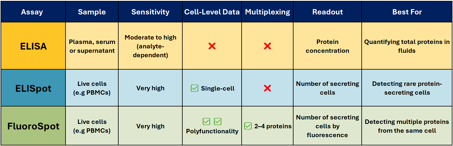
ELISA – The Trusted Workhorse 📊
Best for: Quantifying total soluble protein in biological fluids.
ELISA (Enzyme-Linked Immunosorbent Assay) is one of the most widely used immunoassays for detecting and quantifying soluble proteins, such as cytokines, antibodies, and hormones, in serum, plasma, or cell culture supernatants. It’s robust, scalable, and ideal for high-throughput analysis when you need accurate, reproducible concentration data.
How it works (Sandwich ELISA):
A capture antibody is coated onto a high-binding plate and binds the target protein in your sample. A biotinylated detection antibody binds a different epitope, followed by a streptavidin–enzyme conjugate. Addition of a colourimetric substrate produces a measurable signal proportional to protein concentration.
Why researchers choose it:
- Delivers quantitative results (e.g., pg/ml or ng/ml)
- Compatible with high-throughput and automation
- Well-established, with widely available equipment
- Ideal for comparative analysis across multiple samples
Limitations:
- No information on the number or type of cells producing the protein
- Sensitivity can be lower than single-cell assays
ELISpot – Spotlight on Secreting cells
Best for: Counting individual protein-secreting cells.
ELISpot (Enzyme-Linked ImmunoSpot) detects and counts cells that secrete a specific protein. It’s particularly valuable when studying immune responses where the frequency of antigen-specific T or B cells is low – for example in vaccine research, oncology, and autoimmune disease studies.
How it works:
Live immune cells are added to a PVDF plate pre-coated with a capture antibody. When stimulated, the cells secrete the target protein, which is immediately bound near the cell. After washing away cells, a detection antibody and enzyme conjugate are added. A precipitating substrate forms a visible spot at each secretion site – each spot representing a single responding cell.
Why it stands out:
- Extremely sensitive – can detect one responder cell in >100,000
- Functional readout of immune activity
- Ideal for rare antigen-specific responses
- Widely used in T-cell response monitoring, vaccine trials, allergy research, and immuno-oncology
Pairing with Mabtech’s ASTOR2 automated ELISpot reader delivers rapid, high-precision spot counting with consistent, reproducible results across entire assay plates, and when used with Mabtech monoclonal antibody pairs and ready-to-use ELISpot kits, ensures reliable performance across species and sample types.
FluoroSpot – Multiplex Made Easy 🌈
Best for: Analysing polyfunctional immune responses.
FluoroSpot builds on the ELISpot principle but uses fluorescently labelled detection antibodies to identify multiple proteins secreted by the same cell – typically 2-4 proteins. This allows you to measure not just whether a cell responds, but how it responds.
Why it’s powerful:
- Detects polyfunctionality – cells secreting multiple cytokines
- High sensitivity with low background
- Enables simultaneous detection of different functional subsets
- Excellent for vaccine research, infectious disease, and cancer immunotherapy
Pairing with Mabtech’s IRIS2 ELISpot, FluoroSpot and FociSpot reader enables precise, high-resolution spot detection across multiple fluorescence channels.
Joe’s Takeaway
- ELISA → When you want to know how much protein is present in a sample.
- ELISpot → When you need to know how many cells are producing a protein.
- FluoroSpot → When you need to know which protein a cell is producing, and whether it’s producing more than one at the same time.

In many projects, using ELISA and ELISpot/FluoroSpot together can give the clearest picture — combining quantitative bulk measurements with functional single-cell insights.
If you’d like help selecting the right assay for your research, get in touch with our team at Millennium Science. We’re local experts, and Mabtech’s range covers everything from research use to clinical-grade applications.
Until next time… happy experimenting!
Joe Roberts, PhD
Product Manager
Millennium Science

Paws, Science & Smiles: Millennium Science Bring Your Dog to Work Day
Overview
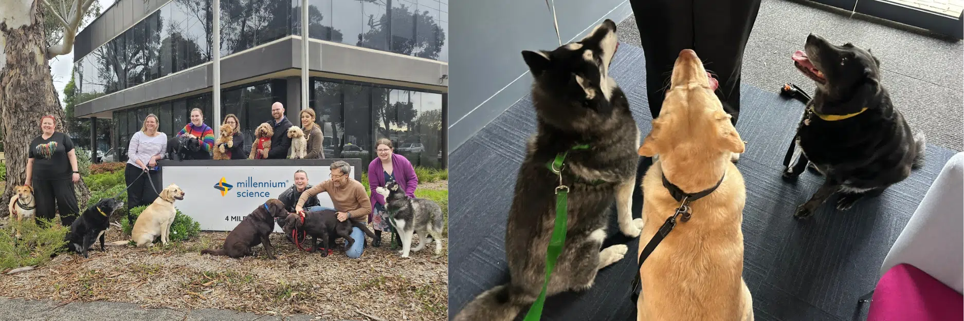
What happens when you mix Labradors, sausage dogs, Irish wolfhounds and a dash of Siberian husky fluff with spreadsheets and science? Magic, apparently. On Tuesday, July 8th, Millennium Science opened its doors (and hearts) to a pack of paws for Bring Your Dog to Work Day, and it was pawsitively 🐾 wonderful. From professional pet portraits to gourmet dog jambalaya, it was a celebration of tails, treats, and top-tier company culture.
Highlights from the Day 🎬
📸 Pet Portraits with a Pro
We kicked things off with a professional pet photographer who captured each pup in their best light. There were solo glamour shots, action shots, and – our crowning achievement – a group photo! Herding cats might be hard, but coordinating a team of dogs to pose together? That’s next-level project management.
🍖 Canine Fine Dining
Lunch was no ordinary kibble situation. Our very own CEO Alex Szabo rolled out his signature dog jambalaya, and it was a gastronomic event for the ages (if you ask the dogs, anyway). Five-star tail wags all round.
🐕 Speed Dating, Dog Style
Let’s face it: dogs invented speed dating. Every hallway stroll turned into a social event. We saw new friendships, unexpected tail-wags, and some solid interspecies networking.
🦴 Treats, Walks & All the Love
Throughout the day, the pups enjoyed scenic walks, an abundance of pats, and doggy bags of treats. Meanwhile, the humans enjoyed what was arguably the most serotonin-boosting Tuesday of the year.
Our Culture
Yes, it was adorable. But Bring Your Dog to Work Day was more than just a flurry of floppy ears and furry faces – it was a reflection of the culture we live every day at Millennium Science.
Dogs are core team members here. Our Company Morale Officers are welcome in the office any day of the week, happily lounging under desks or offering cuddles to anyone. In fact, our official company mascot is none other than Lincoln, our CEO’s beloved chocolate Labrador, and they quite possibly the most popular member of the senior leadership team.
Whether you’re here for the science or the snuggles, there’s no doubt: Millennium Science is a genuinely fantastic place to work.

Pictured: Lincoln Szabo, our company mascot.
Wrapping Up
With 12 dogs, countless treats, one gourmet lunch, and a gallery of professional pet portraits, our Bring Your Dog to Work Day was a howling success, proving once again that the culture at Millennium Science is one-of-a-kind — just like our furry coworkers.
📣 Want to see more of our culture in action?
🐾 Follow us on LinkedIn, BlueSky or X
🚀 Watch our company video
Join the Pack – Careers at Millennium Science Page
On this page
Streamline Microbial Growth Studies with the Agilent BioTek LogPhase 600
Overview
The Agilent BioTek LogPhase™ 600 Microbiology Reader is a high-performance solution for labs studying microbial growth and metabolism. Purpose-built to support real-time, kinetic analysis of bacteria and yeast cultures, the LogPhase 600 enables the simultaneous monitoring of microbial growth curves across up to four standard 96-well microplates – boosting throughput and consistency for microbiology applications.

Precision Temperature Control and Shaking for Reliable Growth Conditions
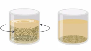
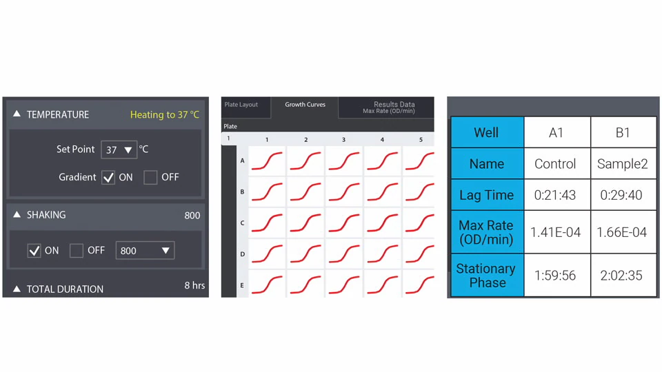
Designed specifically for microbial assays, the LogPhase 600 features robust shaking and sensor-driven temperature control, which are essential for optimal bacterial and yeast cell growth. The integrated orbital shaking mechanism keeps microbial cultures in suspension, promoting uniform growth across all wells.
Advanced temperature regulation ensures uniform heating throughout the instrument, eliminating edge effects and minimising evaporation. A carefully engineered top-to-bottom temperature gradient prevents condensation on sealed plates – reducing light scatter and ensuring accurate optical density (OD) measurements.
Consistent and Reproducible Microbial Growth Curves
Whether you’re performing bacterial growth curve analysis or extended kinetic microbial studies, the LogPhase 600 provides reproducible results across replicates. Its stable environment makes it ideal for long-term microbial monitoring, delivering high-quality data for even the most demanding experimental conditions.
Intuitive Software for Simplified Microbiology Analysis
The LogPhase 600 is controlled via a user-friendly app that simplifies data acquisition and analysis across multiple microplates. Designed with microbiologists in mind, the software allows users to:
-
Start using the instrument with minimal training
-
View multiplate data simultaneously
-
Automatically calculate key growth parameters like Lag time, Maximum growth rate (OD/min) and Time to stationary phase
This makes it easy to integrate the instrument into both academic and industrial lab workflows.
Versatile Applications for High-Throughput Microbiology
With its four-plate capacity, the LogPhase 600 delivers higher throughput than single-plate readers – ideal for labs running large-scale experiments. Common research areas include:
-
Bacterial and yeast growth assays
-
Algal growth studies
-
Antimicrobial resistance research
-
Biofuel production monitoring
-
Food and beverage microbiology testing
Why Choose the Agilent BioTek LogPhase 600?
For researchers seeking a dedicated microbiology plate reader that delivers accuracy, scalability, and ease of use, the LogPhase 600 offers an unmatched combination of throughput, data quality, and application-specific performance. Whether you’re monitoring bacterial growth under various conditions or conducting resistance assays, the LogPhase 600 ensures your data is consistent and actionable.
Contact Us
If you have a question or would like pricing for a LogPhase 600 simply email customerservice@mscience.com.au.
On this page
- Overview
- Precision Temperature Control and Shaking for Reliable Growth Conditions
- Consistent and Reproducible Microbial Growth Curves
- Intuitive Software for Simplified Microbiology Analysis
- Versatile Applications for High-Throughput Microbiology
- Why Choose the Agilent BioTek LogPhase 600?
- Contact Us
In the Lab with Dr Michael Smout
xCELLigence, Hookworms and Wound Healing with Dr Michael Smout. Webinar Recording Now Available!

Recently, Dr Michael Smout from James Cook University, provided an insightful webinar where he delved into his pioneering research on parasitic worms and their potential in advancing wound healing therapies. Central to his studies is the Agilent xCELLigence Real-Time Cell Analysis (RTCA) system, a cutting-edge technology that enables continuous, label-free monitoring of live cells.
Dr. Smout’s work focuses on the granulin protein secreted by parasitic worms, exploring its role in accelerating wound healing—a discovery that holds promise for treating chronic wounds, particularly in diabetic patients.
Webinar Highlights
- Application of xCELLigence RTCA in monitoring cellular responses to parasitic proteins.
- Insights into the therapeutic potential of parasite-derived compounds in wound healing.
- Discussion on the integration of real-time cell analysis in parasitology research.
Agilent xCELLigence RTCA System
The Agilent xCELLigence RTCA system is a state-of-the-art platform that provides real-time, label-free analysis of cellular events. By measuring electrical impedance across microelectrodes integrated into the bottom of specialised plates, the system offers dynamic monitoring of cell proliferation, morphology, and viability. Key features include:
- Continuous, non-invasive monitoring of live cells.
- High sensitivity and reproducibility in detecting cellular changes.
- Versatility across various applications, including cytotoxicity, cell adhesion, and migration assays.
This technology has been instrumental in advancing research across multiple disciplines, providing researchers with a deeper understanding of cellular behaviours in real-time.

Deeper Spatial Discovery with Visium HD 3′
Visium HD 3' is here! Explore more species - Now Shipping
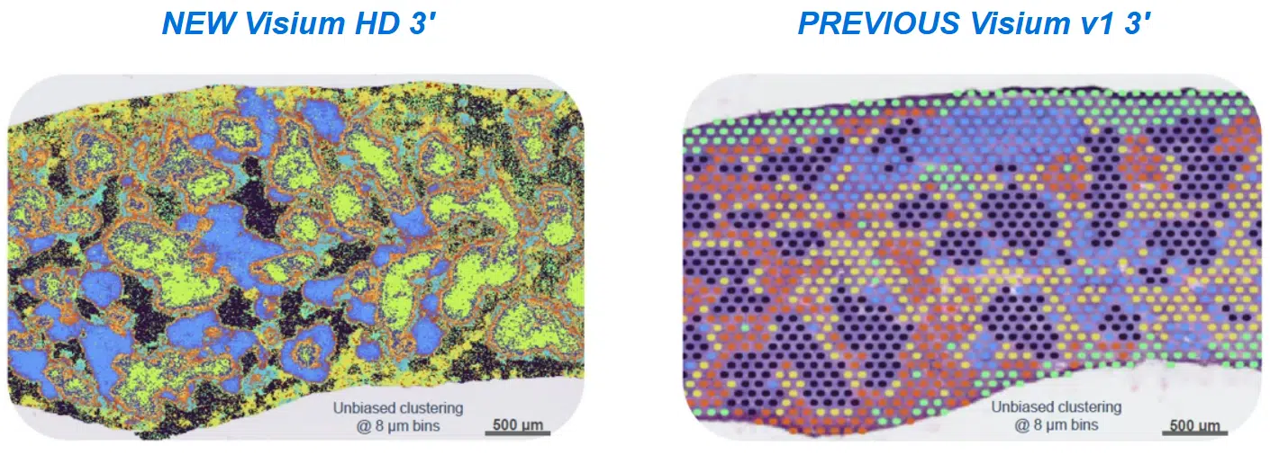
Introducing 10x Genomics‘ Visium HD 3’, a novel assay for unbiased cross-species spatial gene expression profiling of fresh frozen tissue sections mounted on a standard glass slide. The Visium HD’s array has a gapless design that enables integration of unsupervised gene expression clustering data with microscope H&E images from the same tissue section, allowing precise profiling of finer anatomical features with high tissue coverage at single cell-scale resolution.
Furthermore, this novel reverse transcription-based assay generates cDNA products compatible with both short reads and long reads sequencing, extending its applications beyond gene expression analysis to enable immune profiling and the discovery of isoforms and novel transcripts on a spatial level.

Key Features
Visium HD 3’ assay, which delivers whole transcriptome spatial analysis with a 3’ poly(A) capture-based chemistry. This assay provides:
- Flexibility to explore the spatial biology of fresh frozen samples from any species
- De novo spatial discovery power, including isoforms, TCRs/BCRs, and SNVs
- True single cell resolution with new cell segmentation software
- Streamlined slide handling and high spatial fidelity powered by the Visium CytAssist instrument
Space Ranger v4.0.1 is now live! Expand the power of Visium HD with cell segmentation
Improve cell-type resolution with nuclei expansion–based cell segmentation available in Space Ranger version 4.0 software. Importantly, Space Ranger v4.0 now supports cell and nucleus segmentation with H&E images. So you can reprocess existing Visium HD data through the new Space Ranger pipeline to derive these nuclei and cell segmentation outputs alongside the square bins
- Input a brightfield image of your H&E-stained tissue into the Space Ranger pipeline
- The software performs nuclei detection and expansion
- The Space Ranger pipeline outputs custom binning files viewable in Loupe Browser software
The new version can be found at: https://www.10xgenomics.com/support/software/space-ranger/latest

Some other notable features are:
- A new UMI-based registration algorithm for Visium HD and Visium HD 3′ data.
- You can now visualize and analyse Visium HD / Visium HD 3′ cell segmentation in Loupe Browser v9.
Watch introductory webinar on-demand
Learn more about the Visium HD 3’ assay in this introductory webinar, including a Q&A with product experts.

Build your own Visium HD 3’ experiment
Use 10x Genomics’ interactive tool to design your Visium HD 3’ experiment. Answer questions about your sample types and experimental goals, and get kit recommendations.

Other Resources
Press Release: 10x Genomics Expands Visium Platform with Launch of Visium HD 3’ Gene Expression, Unlocking Deeper Spatial Recovery
Grant application resources for Visium products
AACR Annual Meeting (Chicago) Visium HD 3′ poster: Visium HD 3’ enables unbiased whole transcriptome spatial profiling of Tumor microenvironment in fresh frozen cancer tissues at single cell scale resolution.

Revolutionising RNA purification with Automation
Accelerating molecular workflows has never been easier
Norgen Biotek’s Magnetic Bead-Based Total RNA Purification Kit is now fully automated, making high-throughput RNA extraction faster, more reliable, and easier to scale.
Designed to isolate all RNA sizes – including microRNA and small RNA – with exceptional purity and reproducibility, this kit delivers consistent performance across a wide range of sample types. Whether you’re working with tissues, cells, bacteria, viruses, bodily fluids, plants or fungi, Norgen Biotek’s advanced RNA purification technology ensures high-yield results without compromising quality.
Save Time with Automated RNA Purification
Manual RNA extraction can be labour-intensive and prone to variability. Norgen Biotek’s magnetic bead-based kit streamlines the process, reducing hands-on time to just 15 minutes, while enhancing consistency across runs.
Built for Lab Automation Platforms
This kit is fully compatible with leading lab automation platforms, including:
-
Opentrons Flex – a modular and cost-effective automation system designed to scale with your lab.
-
Tecan
-
Thermo Fisher KingFisher
-
Hamilton
-
IsoPure
Seamless integration allows labs to boost throughput, minimise human error, and free up valuable time for high-value analysis and discovery.
Why Choose Norgen Biotek RNA Kits?
✅ Compatible with a broad range of sample types
✅ High-quality RNA including microRNA and small RNA
✅ Automation-ready for reliable scalability
✅ Reduced hands-on time and increased reproducibility
Interested in upgrading your RNA workflows?
Explore how Norgen Biotek and Opentrons can modernise your lab’s RNA extraction today or contact Narisa Dawar our Norgen Biotek Product Manager – ndawar@mscience.com.au.
MedChemExpress Anti-Cancer Compound Library
MedChemExpress (MCE) is a supplier of high-quality chemicals, biochemicals and compound libraries, supporting the global scientific research community. MCE’s extensive product portfolio includes a range of screening libraries designed to accelerate drug discovery and development across multiple therapeutic areas. Among these, the Anti-Cancer Compound Library is a powerful tool for researchers seeking targeted solutions and novel cancer therapies.

The library offers over 8,000 compounds with activity against both solid and haematologic tumours, making it suitable for high-throughput screening (HTS) and high-content screening (HCS).
This collection spans key cancer-related pathways such as apoptosis, cell cycle regulation and signal transduction. It also covers diverse signalling pathways – including kinase, GPCR and epigenetics – providing researchers with a comprehensive platform to explore and identify potential anti-cancer agents.
Many of the compounds have undergone rigorous clinical or preclinical evaluations, with some already approved by the FDA, highlighting their therapeutic potential.
The bioactivity and safety profiles of the compounds make the library a useful resource for advancing cancer research. Each compound is also validated for high purity and quality using NMR and LC/MS techniques, which should lead to precise and reproducible results.
A key advantage of MCE’s compound libraries is that users can fully customise a library to meet research needs from the type, quantity and concentrations of compounds and layout, offering the flexibility that researchers need to drive meaningful discoveries in oncology.
Click here to download the MedChemExpress Compounding Libraries brochure or contact Millennium Science at customerservice@mscience.com.au.
Related Blog
MedChemExpress compound libraries for drug discoveryOn this page
Flow Cytometer detects bacteria in environmental waters
Case Study

The rapid and accurate detection of bacteria in aquatic environments has typically been challenging due to the complexity of the natural water microbial populations. Traditional agar plate methods for analysing aquatic bacteria are subjective and laborious, and rely on being able to adequately culture bacteria.
The good news is, flow cytometers with a high sensitivity of detection provide tools for detecting and analysing microbes independent of their cultivability, as the size, number, nucleic acid content, activity and classification of bacteria can be derived from scattered light and fluorescence signals using flow cytometry. The Agilent NovoCyte flow cytometer can detect very small particles (down to 0.1 µm) with high sensitivity, and so can be applied to various microbial studies by combining multiparameter analytic capability and convenient fluidic maintenance.
In testing the flow cytometer’s capabilities, water samples from multiple environment waters were stained with either SYBR Green I or SYTOX Green I; both are fluorescent dyes which bind to nucleic acids. The instrument was cleaned prior to running samples, filtered buffers were used, and an unstained sample was run to distinguish background noise from fluorescent signal and appropriate scatter and fluorescent thresholding was undertaken.
Using the NovoCyte flow cytometer, the bacteria in natural water could be differentiated into two groups: bacteria of low nucleic acid content (LNA) and of high nucleic acid content (HNA) (Figure 1). It is broadly accepted that HNA is active bacteria, whereas LNA is inactive, dead or a dormant population.

Figure 1: Detection of bacteria in natural waters.
Total bacterial cell counts in multiple environmental waters was also quantified using the NovoCyte flow cytometer (see Table 1). Results demonstrated that natural waters contained the highest bacterial counts of the samples tested and bottled water the lowest. Furthermore, bacterial counts in still water were 10 to 100x higher than that of spring and mountain stream sources.

Table 1: Total cell counts of bacteria in various water samples.
The detection of bacteria in various water samples is essential to maintaining sanitary and healthy drinking conditions, and the NovoCyte flow cytometer can easily and efficiently detect and quantify bacteria in water from several sources. With detection sensitivity coupled to the automatic cell counts measured for each sample, the NovoCyte is an efficient instrument for this application.
Top image credit: iStock.com/stock_colors
This article was initially published in Lab+Life Scientist.
On this page
Expand Your Lab’s Imaging Capabilities with the LICORbio Odyssey F Imaging System
Accelerated Imaging with Exceptional Sensitivity and Speed
The LICORbio Odyssey F Imaging System delivers high sensitivity and broad dynamic range with just a click. Available in 3- and 10-channel models, this advanced platform scans samples up to twice as fast as previous generations, supporting a wide array of applications beyond standard qualitative Western blotting. Whether you’re imaging proteins, nucleic acids, or other biomolecules, the Odyssey F accelerates your workflow without compromising data quality.

Dual NIR and Visible Lasers for Advanced Multiplex Imaging
Equipped with dual near-infrared (NIR) lasers, the Odyssey F ensures optimal throughput and quantitation for high-quality imaging. In addition, two visible fluorescence lasers unlock powerful multiplexing capabilities, enabling researchers to explore multiple targets simultaneously in plate-based assays, slide imaging, and beyond. This flexibility makes it ideal for researchers aiming to capture more data from a single experiment.
Rapid High-Throughput Imaging with Reproducible Results
Users benefit from rapid scan speeds that reduce total imaging time while improving reproducibility. The Odyssey F is engineered for superior sensitivity and consistent performance, giving researchers increased confidence in their data. Its expanded dynamic range means fewer adjustments and more reliable quantification across a broad concentration spectrum.
Versatile Platform for a Wide Range of Imaging Applications
Designed for versatility, the Odyssey F supports a wide variety of assays and sample types. The expanded focus offset range ensures compatibility with numerous plate formats and sample depths. Whether you’re conducting EMSA/Gel Shift Assays, In-Cell Westerns, Tissue Section Imaging, or Whole Slide Imaging (WSI), the Odyssey F adapts to your lab’s needs, enhancing both productivity and application reach.
See the Odyssey F in action!
Book a demoMeet Aunty from Unchained Labs: The Fastest, Highest Throughput Protein Stability Characterisation Platform on the planet!
About Aunty
Unchained Labs has raised the bar in biologics characterisation with the launch of Aunty – the world’s fastest and most high-throughput platform for protein stability analysis. Tailored for researchers in biotech, pharma, and gene therapy, Aunty delivers unprecedented speed, sensitivity, and flexibility across a wide range of stability parameters – all from a single 96-well quartz plate.

Why Aunty is a Game-Changer in Protein and Viral Vector Stability Testing
🔬 Comprehensive Stability Profiling — In Minutes
Aunty characterises protein melting (Tm, Tonset), aggregation (Tagg, Tsize), colloidal stability (kD, B22, G22), long-term stability, and viral vector genome ejection, reading a full 96-well plate every minute. With only 8 µL sample per well, it saves valuable material while delivering maximum insight.
💡 Three Technologies. One Platform.
- Full-Spectrum Fluorescence – For intrinsic or dye-based detection of thermal unfolding.
- Static Light Scattering (SLS) – Detects early-stage aggregation with exceptional sensitivity.
- Dynamic Light Scattering (DLS) – Monitors particle size, polydispersity, and colloidal behaviour.
These modalities can be run in parallel or independently, offering complete flexibility to tailor your assay to the molecule at hand.
Explore Aunty’s Core Applications
1. Thermal Ramp Analysis
Track unfolding and aggregation simultaneously using fluorescence and SLS. Aunty’s sensitivity enables detection down to 25 µg/mL, providing reliable Tm and Tagg measurements across formulations, concentrations, and constructs.
2. Protein and Viral Vector Sizing
Aunty’s ISO-compliant DLS covers particles from 0.3 to 1000 nm. Whether you’re working with therapeutic proteins or viral vectors, it delivers high-precision size measurements and polydispersity profiles in seconds.
3. Colloidal Stability
Quantify aggregation risks under different formulations with Aunty’s automated calculation of kD, B22, and G22. This supports developability assessments for high-concentration biologics and formulation transitions (e.g. IV to SC).
4. Capsid and Genome Stability for AAVs
Using SYBR Gold fluorescence, Aunty detects genome ejection temperatures (Tm) in under an hour. It also pinpoints capsid disruption (Tagg) via SLS – essential for viral vector stability profiling.
5. Isothermal Stability Studies
Run long-term thermal stability tests at set temperatures over hours or days. Monitor changes in unfolding or aggregation over time – with sealed wells for sample integrity and minimal instrument occupation.
The Aunty Plate: Quartz Innovation for Precision Science
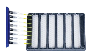
At the heart of Aunty is the first 96-well quartz glass consumable – optimised for superior optical clarity, chemical compatibility, and low-volume use. Automation-friendly and easy to seal, it accelerates workflows and minimises contamination risks.
Fast, Flexible, and Insight-Packed
Aunty’s software simplifies complex workflows with easy setup, real-time monitoring, and intuitive visualisation tools. Whether you’re comparing formulations, screening candidates, or optimising viral vector stability, Aunty delivers actionable results – fast.
Technical Specs at a Glance:
- Sample Volume: 8 µL
- Read Time: 1 minute for 96 samples
- Temperature Range: 15–95°C
- Light Scattering Sensitivity: Down to 0.05 mg/mL (SLS)
- DLS Range: 0.3–1000 nm
- Genome Ejection Sensitivity: ≥5 × 10¹¹ vg/mL
Final Word
Whether you’re optimising a therapeutic antibody, screening excipients, or validating AAV vector stability, Aunty is the gold standard in high-throughput protein and viral vector stability characterisation.
For biotech and pharma researchers looking to speed up developability, reduce material usage, and gain multi-parameter insight in a single run – Aunty is the instrument you’ve been waiting for.
Ready to see Aunty in action?
Contact Millennium Science – your local distributor of Unchained Labs in Australia and New Zealand – for a demo or to request more information.